 Movie
Movie Controller

[English] 日本語
 Yorodumi
- PDB-7ank: Crystal structure of sarcomeric protein FATZ-1 (d91-FATZ-1 constr... -
+ Open data

- Basic information
| Entry | Database: PDB / ID: 7ank | ||||||||||||||||||||||||
|---|---|---|---|---|---|---|---|---|---|---|---|---|---|---|---|---|---|---|---|---|---|---|---|---|---|
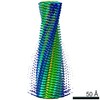
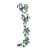
| Title | Crystal structure of sarcomeric protein FATZ-1 (d91-FATZ-1 construct) in complex with half dimer of alpha-actinin-2 | ||||||||||||||||||||||||
 Components Components |
| ||||||||||||||||||||||||
 Keywords Keywords | STRUCTURAL PROTEIN / Z-disk complex / F-actin crosslinking protein / scaffolding protein / fuzzy complex | ||||||||||||||||||||||||
| Function / homology |  Function and homology information Function and homology informationnegative regulation of skeletal muscle tissue regeneration / skeletal muscle fiber adaptation / actin filament uncapping / FATZ binding / titin Z domain binding / positive regulation of endocytic recycling / phospholipase C-activating angiotensin-activated signaling pathway / protein serine/threonine phosphatase inhibitor activity / microspike assembly / telethonin binding ...negative regulation of skeletal muscle tissue regeneration / skeletal muscle fiber adaptation / actin filament uncapping / FATZ binding / titin Z domain binding / positive regulation of endocytic recycling / phospholipase C-activating angiotensin-activated signaling pathway / protein serine/threonine phosphatase inhibitor activity / microspike assembly / telethonin binding / myofibril assembly / negative regulation of protein localization to cell surface / channel activator activity / LIM domain binding / focal adhesion assembly / positive regulation of potassium ion transport / structural constituent of postsynaptic actin cytoskeleton / muscle cell development / cardiac muscle cell development / negative regulation of calcineurin-NFAT signaling cascade / Striated Muscle Contraction / postsynaptic actin cytoskeleton / Nephrin family interactions / actinin binding / Assembly and cell surface presentation of NMDA receptors / sarcomere organization / structural constituent of muscle / cortical actin cytoskeleton / negative regulation of potassium ion transport / Negative regulation of NMDA receptor-mediated neuronal transmission / pseudopodium / Unblocking of NMDA receptors, glutamate binding and activation / skeletal muscle tissue development / Long-term potentiation / postsynaptic density, intracellular component / cytoskeletal protein binding / cell projection / titin binding / phosphatidylinositol-4,5-bisphosphate binding / platelet alpha granule lumen / Ras activation upon Ca2+ influx through NMDA receptor / protein localization to plasma membrane / wound healing / actin filament / filopodium / molecular condensate scaffold activity / regulation of membrane potential / cell junction / postsynaptic density membrane / integrin binding / Z disc / actin filament binding / actin cytoskeleton / Platelet degranulation / RAF/MAP kinase cascade / actin cytoskeleton organization / actin binding / regulation of apoptotic process / dendritic spine / cytoskeleton / transmembrane transporter binding / transcription coactivator activity / cell adhesion / protein domain specific binding / focal adhesion / calcium ion binding / glutamatergic synapse / negative regulation of transcription by RNA polymerase II / extracellular exosome / extracellular region / identical protein binding / nucleus / cytosol Similarity search - Function | ||||||||||||||||||||||||
| Biological species |  Homo sapiens (human) Homo sapiens (human) | ||||||||||||||||||||||||
| Method |  X-RAY DIFFRACTION / SYNCHROTRON / MOLECULAR REPLACEMENT / Resolution: 3.204 Å X-RAY DIFFRACTION / SYNCHROTRON / MOLECULAR REPLACEMENT / Resolution: 3.204 Å | ||||||||||||||||||||||||
 Authors Authors | Sponga, A. / Arolas, J.L. / Rodriguez Chamorro, A. / Mlynek, G. / Hollerl, E. / Schreiner, C. / Pedron, M. / Kostan, J. / Ribeiro, E.A. / Djinovic-Carugo, K. | ||||||||||||||||||||||||
| Funding support |  Austria, Austria,  United Kingdom, 7items United Kingdom, 7items
| ||||||||||||||||||||||||
 Citation Citation | Journal: Sci Adv / Year: 2021 Title: Order from disorder in the sarcomere: FATZ forms a fuzzy but tight complex and phase-separated condensates with alpha-actinin. Authors: Sponga, A. / Arolas, J.L. / Schwarz, T.C. / Jeffries, C.M. / Rodriguez Chamorro, A. / Kostan, J. / Ghisleni, A. / Drepper, F. / Polyansky, A. / De Almeida Ribeiro, E. / Pedron, M. / Zawadzka- ...Authors: Sponga, A. / Arolas, J.L. / Schwarz, T.C. / Jeffries, C.M. / Rodriguez Chamorro, A. / Kostan, J. / Ghisleni, A. / Drepper, F. / Polyansky, A. / De Almeida Ribeiro, E. / Pedron, M. / Zawadzka-Kazimierczuk, A. / Mlynek, G. / Peterbauer, T. / Doto, P. / Schreiner, C. / Hollerl, E. / Mateos, B. / Geist, L. / Faulkner, G. / Kozminski, W. / Svergun, D.I. / Warscheid, B. / Zagrovic, B. / Gautel, M. / Konrat, R. / Djinovic-Carugo, K. | ||||||||||||||||||||||||
| History |
|
- Structure visualization
| Structure viewer | Molecule: MolmilJmol/JSmol |
|---|
- Downloads & links
-Download
| PDBx/mmCIF format | 7ank.cif.gz | 383.8 KB | Display | PDBx/mmCIF format |
|---|---|---|---|---|
| PDB format | pdb7ank.ent.gz | 317.3 KB | Display | PDB format |
| PDBx/mmJSON format | 7ank.json.gz | Tree view | PDBx/mmJSON format | |
| Others |  Other downloads Other downloads |
-Validation report
| Arichive directory | https://data.pdbj.org/pub/pdb/validation_reports/an/7ankftp://data.pdbj.org/pub/pdb/validation_reports/an/7ank | HTTPS FTP |
|---|
-Related structure data
| Related structure data |  7a8tC  7a8uC  4d1eS C: citing same article ( S: Starting model for refinement |
|---|---|
| Similar structure data | |
| Other databases |
|
-Links
 PDBj
PDBj


- Assembly
| Deposited unit | 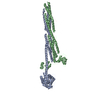
| ||||||||
|---|---|---|---|---|---|---|---|---|---|
| 1 |
| ||||||||
| Unit cell |
|
-Components
| #1: Protein | Mass: 62561.102 Da / Num. of mol.: 1 Source method: isolated from a genetically manipulated source Source: (gene. exp.) Homo sapiens (human) / Gene: ACTN2 / Plasmid: modified pET-8 vector / Production host:  |
|---|---|
| #2: Protein | Mass: 44608.980 Da / Num. of mol.: 1 Source method: isolated from a genetically manipulated source Source: (gene. exp.) Homo sapiens (human) / Gene: ACTN2 / Plasmid: modified pET-8 vector / Production host: |
| #3: Protein | Mass: 21524.656 Da / Num. of mol.: 1 Source method: isolated from a genetically manipulated source Details: UNP Q9NP98 Sequence start 92 Sequence end 299 / Source: (gene. exp.) Homo sapiens (human) / Gene: MYOZ1, MYOZ / Plasmid: modified pET-46 vector / Production host: |
-Experimental details
-Experiment
| Experiment | Method: X-RAY DIFFRACTION / Number of used crystals: 1 |
|---|
- Sample preparation
| Crystal | Density Matthews: 3.16 Å3/Da / Density % sol: 61.1 % |
|---|---|
| Crystal grow | Temperature: 277 K / Method: vapor diffusion, hanging drop / pH: 7.5 Details: 100 mM Bis-Tris propane (pH 7.5), 100 mM sodium citrate, 10 mM zinc chloride, 12% w/w polyethylene glycol 3,350 |
-Data collection
| Diffraction | Mean temperature: 100 K / Serial crystal experiment: N |
|---|---|
| Diffraction source | Source: SYNCHROTRON / Site: ESRF  / Beamline: ID29 / Wavelength: 0.976251 Å / Beamline: ID29 / Wavelength: 0.976251 Å |
| Detector | Type: DECTRIS PILATUS 6M / Detector: PIXEL / Date: Feb 12, 2017 |
| Radiation | Protocol: SINGLE WAVELENGTH / Monochromatic (M) / Laue (L): M / Scattering type: x-ray |
| Radiation wavelength | Wavelength: 0.976251 Å / Relative weight: 1 |
| Reflection | Resolution: 3.2→46.9 Å / Num. obs: 26764 / % possible obs: 98.9 % / Redundancy: 6.6 % / Biso Wilson estimate: 127.5 Å2 / CC1/2: 0.998 / Rmerge(I) obs: 0.097 / Net I/σ(I): 14.1 |
| Reflection shell | Resolution: 3.2→3.29 Å / Redundancy: 6 % / Rmerge(I) obs: 1.425 / Mean I/σ(I) obs: 1.4 / Num. unique obs: 1897 / CC1/2: 0.872 / % possible all: 93.9 |
- Processing
| Software |
| ||||||||||||||||||||||||||||||||||||||||||||||||||||||||||||||||||||||||||||||||||||||||||||||||||||||||||||||||||||||||||||||||||||||||||||||||||||||||||||||||||||||||||||||||||||||||||||||||||||||||||||||||||||||||||||||||||||||||||||||||||||||||||||||||||||||||||||||||||||||||||||||||||||||||||||
|---|---|---|---|---|---|---|---|---|---|---|---|---|---|---|---|---|---|---|---|---|---|---|---|---|---|---|---|---|---|---|---|---|---|---|---|---|---|---|---|---|---|---|---|---|---|---|---|---|---|---|---|---|---|---|---|---|---|---|---|---|---|---|---|---|---|---|---|---|---|---|---|---|---|---|---|---|---|---|---|---|---|---|---|---|---|---|---|---|---|---|---|---|---|---|---|---|---|---|---|---|---|---|---|---|---|---|---|---|---|---|---|---|---|---|---|---|---|---|---|---|---|---|---|---|---|---|---|---|---|---|---|---|---|---|---|---|---|---|---|---|---|---|---|---|---|---|---|---|---|---|---|---|---|---|---|---|---|---|---|---|---|---|---|---|---|---|---|---|---|---|---|---|---|---|---|---|---|---|---|---|---|---|---|---|---|---|---|---|---|---|---|---|---|---|---|---|---|---|---|---|---|---|---|---|---|---|---|---|---|---|---|---|---|---|---|---|---|---|---|---|---|---|---|---|---|---|---|---|---|---|---|---|---|---|---|---|---|---|---|---|---|---|---|---|---|---|---|---|---|---|---|---|---|---|---|---|---|---|---|---|---|---|---|---|---|---|---|---|---|---|---|---|---|---|---|---|---|---|---|---|---|---|---|---|---|---|---|---|---|---|---|---|---|---|---|---|---|---|---|---|---|
| Refinement | Method to determine structure: MOLECULAR REPLACEMENT Starting model: 4D1E Resolution: 3.204→46.94 Å / Cor.coef. Fo:Fc: 0.899 / Cor.coef. Fo:Fc free: 0.89 / Cross valid method: THROUGHOUT / SU Rfree Blow DPI: 0.462
| ||||||||||||||||||||||||||||||||||||||||||||||||||||||||||||||||||||||||||||||||||||||||||||||||||||||||||||||||||||||||||||||||||||||||||||||||||||||||||||||||||||||||||||||||||||||||||||||||||||||||||||||||||||||||||||||||||||||||||||||||||||||||||||||||||||||||||||||||||||||||||||||||||||||||||||
| Displacement parameters | Biso mean: 206.61 Å2
| ||||||||||||||||||||||||||||||||||||||||||||||||||||||||||||||||||||||||||||||||||||||||||||||||||||||||||||||||||||||||||||||||||||||||||||||||||||||||||||||||||||||||||||||||||||||||||||||||||||||||||||||||||||||||||||||||||||||||||||||||||||||||||||||||||||||||||||||||||||||||||||||||||||||||||||
| Refine analyze | Luzzati coordinate error obs: 0.68 Å | ||||||||||||||||||||||||||||||||||||||||||||||||||||||||||||||||||||||||||||||||||||||||||||||||||||||||||||||||||||||||||||||||||||||||||||||||||||||||||||||||||||||||||||||||||||||||||||||||||||||||||||||||||||||||||||||||||||||||||||||||||||||||||||||||||||||||||||||||||||||||||||||||||||||||||||
| Refinement step | Cycle: LAST / Resolution: 3.204→46.94 Å
| ||||||||||||||||||||||||||||||||||||||||||||||||||||||||||||||||||||||||||||||||||||||||||||||||||||||||||||||||||||||||||||||||||||||||||||||||||||||||||||||||||||||||||||||||||||||||||||||||||||||||||||||||||||||||||||||||||||||||||||||||||||||||||||||||||||||||||||||||||||||||||||||||||||||||||||
| Refine LS restraints |
| ||||||||||||||||||||||||||||||||||||||||||||||||||||||||||||||||||||||||||||||||||||||||||||||||||||||||||||||||||||||||||||||||||||||||||||||||||||||||||||||||||||||||||||||||||||||||||||||||||||||||||||||||||||||||||||||||||||||||||||||||||||||||||||||||||||||||||||||||||||||||||||||||||||||||||||
| LS refinement shell | Resolution: 3.204→3.23 Å
| ||||||||||||||||||||||||||||||||||||||||||||||||||||||||||||||||||||||||||||||||||||||||||||||||||||||||||||||||||||||||||||||||||||||||||||||||||||||||||||||||||||||||||||||||||||||||||||||||||||||||||||||||||||||||||||||||||||||||||||||||||||||||||||||||||||||||||||||||||||||||||||||||||||||||||||
| Refinement TLS params. | Refine-ID: X-RAY DIFFRACTION
| ||||||||||||||||||||||||||||||||||||||||||||||||||||||||||||||||||||||||||||||||||||||||||||||||||||||||||||||||||||||||||||||||||||||||||||||||||||||||||||||||||||||||||||||||||||||||||||||||||||||||||||||||||||||||||||||||||||||||||||||||||||||||||||||||||||||||||||||||||||||||||||||||||||||||||||
| Refinement TLS group |
|
