 Movie
Movie Controller


+ Open data

- Basic information
| Entry | Database: PDB / ID: 7akv | |||||||||||||||
|---|---|---|---|---|---|---|---|---|---|---|---|---|---|---|---|---|
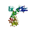
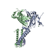
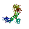
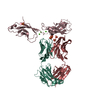
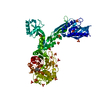
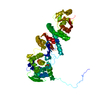
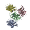
| Title | The cryo-EM structure of the Vag8-C1 inhibitor complex | |||||||||||||||
 Components Components |
| |||||||||||||||
 Keywords Keywords | PROTEIN BINDING / complex bacterial protein human protein immune system inhibitor | |||||||||||||||
| Function / homology |  Function and homology information Function and homology informationnegative regulation of complement activation, lectin pathway / Defective SERPING1 causes hereditary angioedema / outer membrane / blood circulation / fibrinolysis / : / platelet alpha granule lumen / Regulation of Complement cascade / serine-type endopeptidase inhibitor activity / blood coagulation ...negative regulation of complement activation, lectin pathway / Defective SERPING1 causes hereditary angioedema / outer membrane / blood circulation / fibrinolysis / : / platelet alpha granule lumen / Regulation of Complement cascade / serine-type endopeptidase inhibitor activity / blood coagulation / Platelet degranulation / extracellular matrix / blood microparticle / endoplasmic reticulum lumen / : / extracellular exosome / extracellular region Similarity search - Function | |||||||||||||||
| Biological species |  Homo sapiens (human) Homo sapiens (human) Bordetella pertussis (bacteria) Bordetella pertussis (bacteria) | |||||||||||||||
| Method | ELECTRON MICROSCOPY / single particle reconstruction / cryo EM / Resolution: 3.6 Å | |||||||||||||||
 Authors Authors | Johnson, S. / Lea, S.M. / Deme, J.C. / Furlong, E. / Dhillon, A. | |||||||||||||||
| Funding support |  United Kingdom, 4items United Kingdom, 4items
| |||||||||||||||
 Citation Citation | Journal: mBio / Year: 2021 Title: Molecular Basis for Bordetella pertussis Interference with Complement, Coagulation, Fibrinolytic, and Contact Activation Systems: the Cryo-EM Structure of the Vag8-C1 Inhibitor Complex. Authors: Arun Dhillon / Justin C Deme / Emily Furlong / Dorina Roem / Ilse Jongerius / Steven Johnson / Susan M Lea /  Abstract: Complement, contact activation, coagulation, and fibrinolysis are serum protein cascades that need strict regulation to maintain human health. Serum glycoprotein, a C1 inhibitor (C1-INH), is a key ...Complement, contact activation, coagulation, and fibrinolysis are serum protein cascades that need strict regulation to maintain human health. Serum glycoprotein, a C1 inhibitor (C1-INH), is a key regulator (inhibitor) of serine proteases of all the above-mentioned pathways. Recently, an autotransporter protein, virulence-associated gene 8 (Vag8), produced by the whooping cough pathogen, , was shown to bind to C1-INH and interfere with its function. Here, we present the structure of the Vag8-C1-INH complex determined using cryo-electron microscopy at a 3.6-Å resolution. The structure shows a unique mechanism of C1-INH inhibition not employed by other pathogens, where Vag8 sequesters the reactive center loop of C1-INH, preventing its interaction with the target proteases. The structure of a 10-kDa protein complex is one of the smallest to be determined using cryo-electron microscopy at high resolution. The structure reveals that C1-INH is sequestered in an inactivated state by burial of the reactive center loop in Vag8. By so doing, the bacterium is able to simultaneously perturb the many pathways regulated by C1-INH. Virulence mechanisms such as the one described here assume more importance given the emerging evidence about dysregulation of contact activation, coagulation, and fibrinolysis leading to COVID-19 pneumonia. | |||||||||||||||
| History |
|
- Structure visualization
| Movie |
Movie viewer |
|---|---|
| Structure viewer | Molecule: MolmilJmol/JSmol |
- Downloads & links
-Download
| PDBx/mmCIF format | 7akv.cif.gz | 164.7 KB | Display | PDBx/mmCIF format |
|---|---|---|---|---|
| PDB format | pdb7akv.ent.gz | 118.2 KB | Display | PDB format |
| PDBx/mmJSON format | 7akv.json.gz | Tree view | PDBx/mmJSON format | |
| Others |  Other downloads Other downloads |
-Validation report
| Arichive directory | https://data.pdbj.org/pub/pdb/validation_reports/ak/7akvftp://data.pdbj.org/pub/pdb/validation_reports/ak/7akv | HTTPS FTP |
|---|
-Related structure data
| Related structure data |  11814MC M: map data used to model this data C: citing same article ( |
|---|---|
| Similar structure data |

-Links
 PDBj
PDBj


- Assembly
| Deposited unit | 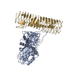
|
|---|---|
| 1 |
|
-Components
| #1: Protein | Mass: 45011.637 Da / Num. of mol.: 1 Source method: isolated from a genetically manipulated source Source: (gene. exp.) Homo sapiens (human) / Gene: SERPING1, C1IN, C1NH / Production host:  |
|---|---|
| #2: Protein | Mass: 91262.172 Da / Num. of mol.: 1 Source method: isolated from a genetically manipulated source Source: (gene. exp.) Bordetella pertussis (bacteria) / Gene: vag-8 / Production host: |
| Has protein modification | Y |
-Experimental details
-Experiment
| Experiment | Method: ELECTRON MICROSCOPY |
|---|---|
| EM experiment | Aggregation state: PARTICLE / 3D reconstruction method: single particle reconstruction |
- Sample preparation
| Component | Name: Vag8:C1 inhibitor complex / Type: COMPLEX / Entity ID: all / Source: MULTIPLE SOURCES |
|---|---|
| Molecular weight | Value: 0.1 MDa / Experimental value: NO |
| Buffer solution | pH: 8 |
| Specimen | Embedding applied: NO / Shadowing applied: NO / Staining applied: NO / Vitrification applied: YES |
| Vitrification | Cryogen name: ETHANE |
- Electron microscopy imaging
| Experimental equipment |  Model: Titan Krios / Image courtesy: FEI Company |
|---|---|
| Microscopy | Model: FEI TITAN KRIOS |
| Electron gun | Electron source:  FIELD EMISSION GUN / Accelerating voltage: 300 kV / Illumination mode: FLOOD BEAM FIELD EMISSION GUN / Accelerating voltage: 300 kV / Illumination mode: FLOOD BEAM |
| Electron lens | Mode: BRIGHT FIELD |
| Image recording | Electron dose: 60 e/Å2 / Film or detector model: GATAN K3 BIOQUANTUM (6k x 4k) |
- Processing
| Software |
| ||||||||||||||||||||||||||||
|---|---|---|---|---|---|---|---|---|---|---|---|---|---|---|---|---|---|---|---|---|---|---|---|---|---|---|---|---|---|
| EM software |
| ||||||||||||||||||||||||||||
| CTF correction | Type: PHASE FLIPPING AND AMPLITUDE CORRECTION | ||||||||||||||||||||||||||||
| Particle selection | Num. of particles selected: 17261358 | ||||||||||||||||||||||||||||
| Symmetry | Point symmetry: C1 (asymmetric) | ||||||||||||||||||||||||||||
| 3D reconstruction | Resolution: 3.6 Å / Resolution method: FSC 0.143 CUT-OFF / Num. of particles: 687883 / Symmetry type: POINT | ||||||||||||||||||||||||||||
| Atomic model building | Protocol: OTHER / Space: REAL | ||||||||||||||||||||||||||||
| Refinement | Cross valid method: NONE Stereochemistry target values: GeoStd + Monomer Library + CDL v1.2 | ||||||||||||||||||||||||||||
| Displacement parameters | Biso mean: 103.34 Å2 | ||||||||||||||||||||||||||||
| Refine LS restraints |
|
