Journal: mBio / Year: 2021 Title: Molecular Basis for Bordetella pertussis Interference with Complement, Coagulation, Fibrinolytic, and Contact Activation Systems: the Cryo-EM Structure of the Vag8-C1 Inhibitor Complex. Authors: Arun Dhillon / Justin C Deme / Emily Furlong / Dorina Roem / Ilse Jongerius / Steven Johnson / Susan M Lea / Abstract: Complement, contact activation, coagulation, and fibrinolysis are serum protein cascades that need strict regulation to maintain human health. Serum glycoprotein, a C1 inhibitor (C1-INH), is a key ...Complement, contact activation, coagulation, and fibrinolysis are serum protein cascades that need strict regulation to maintain human health. Serum glycoprotein, a C1 inhibitor (C1-INH), is a key regulator (inhibitor) of serine proteases of all the above-mentioned pathways. Recently, an autotransporter protein, virulence-associated gene 8 (Vag8), produced by the whooping cough pathogen, , was shown to bind to C1-INH and interfere with its function. Here, we present the structure of the Vag8-C1-INH complex determined using cryo-electron microscopy at a 3.6-Å resolution. The structure shows a unique mechanism of C1-INH inhibition not employed by other pathogens, where Vag8 sequesters the reactive center loop of C1-INH, preventing its interaction with the target proteases. The structure of a 10-kDa protein complex is one of the smallest to be determined using cryo-electron microscopy at high resolution. The structure reveals that C1-INH is sequestered in an inactivated state by burial of the reactive center loop in Vag8. By so doing, the bacterium is able to simultaneously perturb the many pathways regulated by C1-INH. Virulence mechanisms such as the one described here assume more importance given the emerging evidence about dysregulation of contact activation, coagulation, and fibrinolysis leading to COVID-19 pneumonia.
History
Deposition
Oct 2, 2020
-
Header (metadata) release
Jun 16, 2021
-
Map release
Jun 16, 2021
-
Update
Oct 23, 2024
-
Current status
Oct 23, 2024
Processing site: PDBe / Status: Released
-
Structure visualization
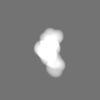
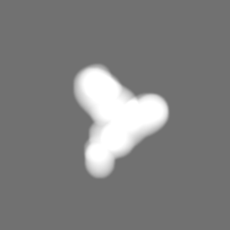
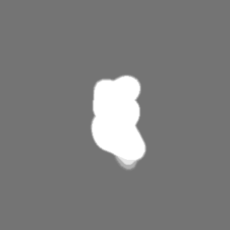
Movie
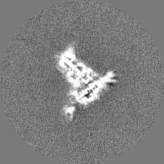
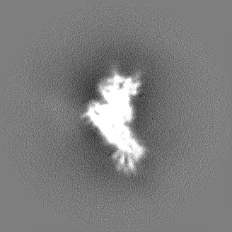
Surface view with section colored by density value
In the structure databanks used in Yorodumi, some data are registered as the other names, "COVID-19 virus" and "2019-nCoV". Here are the details of the virus and the list of structure data.
Jan 31, 2019. EMDB accession codes are about to change! (news from PDBe EMDB page)
EMDB accession codes are about to change! (news from PDBe EMDB page)
The allocation of 4 digits for EMDB accession codes will soon come to an end. Whilst these codes will remain in use, new EMDB accession codes will include an additional digit and will expand incrementally as the available range of codes is exhausted. The current 4-digit format prefixed with “EMD-” (i.e. EMD-XXXX) will advance to a 5-digit format (i.e. EMD-XXXXX), and so on. It is currently estimated that the 4-digit codes will be depleted around Spring 2019, at which point the 5-digit format will come into force.
The EM Navigator/Yorodumi systems omit the EMD- prefix.
Related info.:Q: What is EMD? / ID/Accession-code notation in Yorodumi/EM Navigator
Yorodumi is a browser for structure data from EMDB, PDB, SASBDB, etc.
This page is also the successor to EM Navigator detail page, and also detail information page/front-end page for Omokage search.
The word "yorodu" (or yorozu) is an old Japanese word meaning "ten thousand". "mi" (miru) is to see.
Related info.:EMDB / PDB / SASBDB / Comparison of 3 databanks / Yorodumi Search / Aug 31, 2016. New EM Navigator & Yorodumi / Yorodumi Papers / Jmol/JSmol / Function and homology information / Changes in new EM Navigator and Yorodumi
 Movie
Movie Controller


 Open data
 Basic information
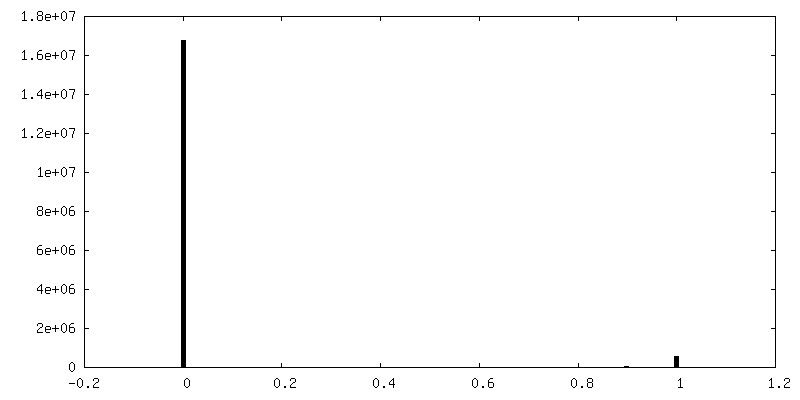
 Map data
 Sample
 Keywords
 Function and homology information
 Homo sapiens (human) /
  Bordetella pertussis (bacteria)
 Authors
 United Kingdom, 4 items
  Citation
 Structure visualization
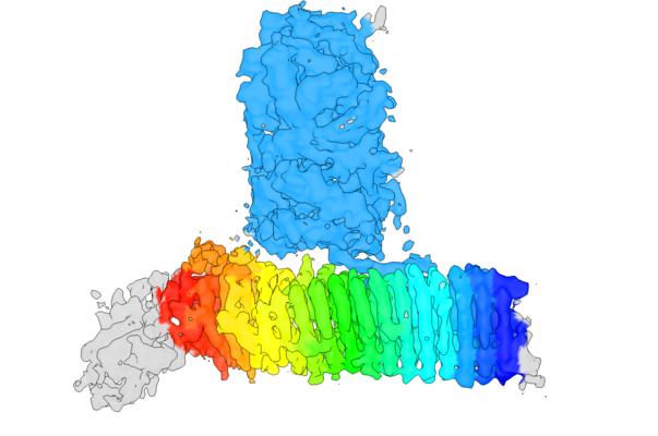
 Downloads & links
 emd_11814.png
 http://ftp.pdbj.org/pub/emdb/structures/EMD-11814


 Z (Sec.)
 Y (Row.)
 X (Col.)


 Sample components
 Processing
 Electron microscopy
 FIELD EMISSION GUN

