 Movie
Movie Controller

[English] 日本語
 Yorodumi
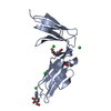
- PDB-3luo: Crystal Structure and functional characterization of the thermoph... -
+ Open data

- Basic information
| Entry | Database: PDB / ID: 3luo | ||||||
|---|---|---|---|---|---|---|---|
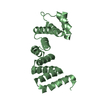
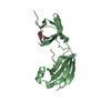
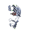
| Title | Crystal Structure and functional characterization of the thermophilic prolyl isomerase and chaperone SlyD | ||||||
 Components Components |
| ||||||
 Keywords Keywords | ISOMERASE / PROLYL CIS TRANS ISOMERASE / CHAPERONE FUNCTION / TWO DOMAIN PROTEIN / Ni(2+) Zn(2+) BINDING / SLYD | ||||||
| Function / homology |  Function and homology information Function and homology informationpeptidylprolyl isomerase / peptidyl-prolyl cis-trans isomerase activity / protein refolding / metal ion binding / cytoplasm Similarity search - Function | ||||||
| Biological species |   Thermus thermophilus (bacteria) Thermus thermophilus (bacteria) | ||||||
| Method |  X-RAY DIFFRACTION / SYNCHROTRON / FOURIER SYNTHESIS / Resolution: 2.55 Å X-RAY DIFFRACTION / SYNCHROTRON / FOURIER SYNTHESIS / Resolution: 2.55 Å | ||||||
 Authors Authors | Loew, C. / Neumann, P. / Weininger, U. / Stubbs, M.T. / Balbach, J. | ||||||
 Citation Citation | Journal: J.Mol.Biol. / Year: 2010 Title: Crystal Structure Determination and Functional Characterization of the Metallochaperone SlyD from Thermus thermophilus Authors: Loew, C. / Neumann, P. / Tidow, H. / Weininger, U. / Haupt, C. / Friedrich-Epler, B. / Scholz, C. / Stubbs, M.T. / Balbach, J. | ||||||
| History |
|
- Structure visualization
| Structure viewer | Molecule: MolmilJmol/JSmol |
|---|
- Downloads & links
-Download
| PDBx/mmCIF format | 3luo.cif.gz | 75.9 KB | Display | PDBx/mmCIF format |
|---|---|---|---|---|
| PDB format | pdb3luo.ent.gz | 56.5 KB | Display | PDB format |
| PDBx/mmJSON format | 3luo.json.gz | Tree view | PDBx/mmJSON format | |
| Others |  Other downloads Other downloads |
-Validation report
| Arichive directory | https://data.pdbj.org/pub/pdb/validation_reports/lu/3luoftp://data.pdbj.org/pub/pdb/validation_reports/lu/3luo | HTTPS FTP |
|---|
-Related structure data
| Related structure data |  3cgmSC 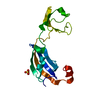 3cgnC S: Starting model for refinement C: citing same article ( |
|---|---|
| Similar structure data |


-Links
 PDBj
PDBj

- Assembly
| Deposited unit | 
| ||||||||
|---|---|---|---|---|---|---|---|---|---|
| 1 |
| ||||||||
| 2 | 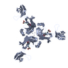
| ||||||||
| Unit cell |
|
-Components
| #1: Protein | Mass: 17400.234 Da / Num. of mol.: 1 Source method: isolated from a genetically manipulated source Source: (gene. exp.) Thermus thermophilus (bacteria) / Strain: HB8 / Plasmid: PET-VECTOR / Production host: | ||
|---|---|---|---|
| #2: Protein/peptide | Mass: 590.625 Da / Num. of mol.: 1 / Source method: obtained synthetically Details: THE PEPTIDE SUBSTRATE WAS PURCHASED FROM A COMPANY. | ||
| #3: Chemical | ChemComp-ZN /   Mass: 65.409 Da / Num. of mol.: 4 / Source method: obtained synthetically / Formula: Zn Mass: 65.409 Da / Num. of mol.: 4 / Source method: obtained synthetically / Formula: Zn#4: Water | ChemComp-HOH / |  Mass: 18.015 Da / Num. of mol.: 21 / Source method: isolated from a natural source / Formula: H2O Mass: 18.015 Da / Num. of mol.: 21 / Source method: isolated from a natural source / Formula: H2O |
-Experimental details
-Experiment
| Experiment | Method: X-RAY DIFFRACTION / Number of used crystals: 1 |
|---|
- Sample preparation
| Crystal | Density Matthews: 3.2 Å3/Da / Density % sol: 61.58 % |
|---|---|
| Crystal grow | Temperature: 298 K / Method: vapor diffusion, hanging drop / pH: 6.5 Details: 10MG/ML N-SUC-ALA-LEU-PRO-PHE-PNA, 2M AMMONIUM SULFATE, 5 % PEG 400, 100MM MES, PH 6.5, VAPOR DIFFUSION, HANGING DROP, temperature 298K |
-Data collection
| Diffraction | Mean temperature: 100 K |
|---|---|
| Diffraction source | Source: SYNCHROTRON / Site: BESSY  / Beamline: 14.1 / Wavelength: 0.9841 Å / Beamline: 14.1 / Wavelength: 0.9841 Å |
| Detector | Type: MARMOSAIC 225 mm CCD / Detector: CCD / Date: Jun 12, 2009 / Details: mirrors |
| Radiation | Monochromator: GRAPHITE / Protocol: SINGLE WAVELENGTH / Monochromatic (M) / Laue (L): M / Scattering type: x-ray |
| Radiation wavelength | Wavelength: 0.9841 Å / Relative weight: 1 |
| Reflection | Resolution: 2.55→20 Å / Num. all: 7658 / Num. obs: 7636 / % possible obs: 99.7 % / Observed criterion σ(F): 0 / Observed criterion σ(I): -3 / Redundancy: 7.35 % / Biso Wilson estimate: 68.806 Å2 / Rmerge(I) obs: 0.05 / Rsym value: 0.054 / Net I/σ(I): 23.77 |
| Reflection shell | Resolution: 2.55→2.65 Å / Redundancy: 7.2 % / Rmerge(I) obs: 0.643 / Mean I/σ(I) obs: 3.5 / Num. measured obs: 5982 / Num. unique all: 808 / Num. unique obs: 808 / Rsym value: 0.662 / % possible all: 100 |
- Processing
| Software |
| ||||||||||||||||||||||||||||||||||||||||||||||||||||||||||||||||||||||||||||||||||||||||||||||||||||||||||||||||
|---|---|---|---|---|---|---|---|---|---|---|---|---|---|---|---|---|---|---|---|---|---|---|---|---|---|---|---|---|---|---|---|---|---|---|---|---|---|---|---|---|---|---|---|---|---|---|---|---|---|---|---|---|---|---|---|---|---|---|---|---|---|---|---|---|---|---|---|---|---|---|---|---|---|---|---|---|---|---|---|---|---|---|---|---|---|---|---|---|---|---|---|---|---|---|---|---|---|---|---|---|---|---|---|---|---|---|---|---|---|---|---|---|---|
| Refinement | Method to determine structure: FOURIER SYNTHESIS Starting model: PDB ENTRY 3CGM Resolution: 2.55→19.693 Å / Occupancy max: 1 / Occupancy min: 0.43 / SU ML: 0.33 / Isotropic thermal model: Isotropic / Cross valid method: THROUGHOUT / σ(F): 1.24 / σ(I): 0 / Stereochemistry target values: ML / Details: TLS parameters have been included in refinemnt
| ||||||||||||||||||||||||||||||||||||||||||||||||||||||||||||||||||||||||||||||||||||||||||||||||||||||||||||||||
| Solvent computation | Shrinkage radii: 0.8 Å / VDW probe radii: 0.8 Å / Solvent model: FLAT BULK SOLVENT MODEL / Bsol: 92.506 Å2 / ksol: 0.407 e/Å3 | ||||||||||||||||||||||||||||||||||||||||||||||||||||||||||||||||||||||||||||||||||||||||||||||||||||||||||||||||
| Displacement parameters | Biso max: 215.89 Å2 / Biso mean: 94.308 Å2 / Biso min: 31.23 Å2
| ||||||||||||||||||||||||||||||||||||||||||||||||||||||||||||||||||||||||||||||||||||||||||||||||||||||||||||||||
| Refine analyze |
| ||||||||||||||||||||||||||||||||||||||||||||||||||||||||||||||||||||||||||||||||||||||||||||||||||||||||||||||||
| Refinement step | Cycle: LAST / Resolution: 2.55→19.693 Å
| ||||||||||||||||||||||||||||||||||||||||||||||||||||||||||||||||||||||||||||||||||||||||||||||||||||||||||||||||
| Refine LS restraints |
| ||||||||||||||||||||||||||||||||||||||||||||||||||||||||||||||||||||||||||||||||||||||||||||||||||||||||||||||||
| LS refinement shell | Refine-ID: X-RAY DIFFRACTION / Total num. of bins used: 15
| ||||||||||||||||||||||||||||||||||||||||||||||||||||||||||||||||||||||||||||||||||||||||||||||||||||||||||||||||
| Refinement TLS params. | Method: refined / Refine-ID: X-RAY DIFFRACTION
| ||||||||||||||||||||||||||||||||||||||||||||||||||||||||||||||||||||||||||||||||||||||||||||||||||||||||||||||||
| Refinement TLS group |
|
