 Movie
Movie Controller


+ Open data

- Basic information
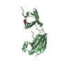
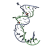
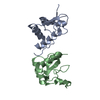
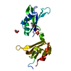
| Entry | Database: PDB / ID: 3cgn | ||||||
|---|---|---|---|---|---|---|---|
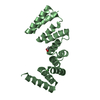
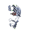
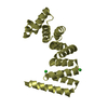
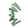
| Title | Crystal Structure of thermophilic SlyD | ||||||
 Components Components | Peptidyl-prolyl cis-trans isomerase | ||||||
 Keywords Keywords | ISOMERASE / SlyD / prolyl cis trans isomerase / chaperone / Rotamase | ||||||
| Function / homology |  Function and homology information Function and homology informationpeptidylprolyl isomerase / peptidyl-prolyl cis-trans isomerase activity / protein refolding / metal ion binding / cytoplasm Similarity search - Function | ||||||
| Biological species |   Thermus thermophilus (bacteria) Thermus thermophilus (bacteria) | ||||||
| Method |  X-RAY DIFFRACTION / SYNCHROTRON / MOLECULAR REPLACEMENT / molecular replacement / Resolution: 2.7 Å X-RAY DIFFRACTION / SYNCHROTRON / MOLECULAR REPLACEMENT / molecular replacement / Resolution: 2.7 Å | ||||||
 Authors Authors | Neumann, P. / Loew, C. / Stubbs, M.T. / Balbach, J. | ||||||
 Citation Citation | Journal: J.Mol.Biol. / Year: 2010 Title: Crystal Structure Determination and Functional Characterization of the Metallochaperone SlyD from Thermus thermophilus Authors: Loew, C. / Neumann, P. / Tidow, H. / Weininger, U. / Haupt, C. / Friedrich-Epler, B. / Scholz, C. / Stubbs, M.T. / Balbach, J. | ||||||
| History |
|
- Structure visualization
| Structure viewer | Molecule: MolmilJmol/JSmol |
|---|
- Downloads & links
-Download
| PDBx/mmCIF format | 3cgn.cif.gz | 43 KB | Display | PDBx/mmCIF format |
|---|---|---|---|---|
| PDB format | pdb3cgn.ent.gz | 29.6 KB | Display | PDB format |
| PDBx/mmJSON format | 3cgn.json.gz | Tree view | PDBx/mmJSON format | |
| Others |  Other downloads Other downloads |
-Validation report
| Arichive directory | https://data.pdbj.org/pub/pdb/validation_reports/cg/3cgnftp://data.pdbj.org/pub/pdb/validation_reports/cg/3cgn | HTTPS FTP |
|---|
-Related structure data
| Related structure data |  3cgmSC  3luoC S: Starting model for refinement C: citing same article ( |
|---|---|
| Similar structure data |


-Links
 PDBj
PDBj


- Assembly
| Deposited unit | 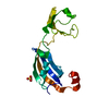
| ||||||||
|---|---|---|---|---|---|---|---|---|---|
| 1 |
| ||||||||
| 2 | 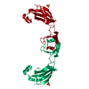
| ||||||||
| Unit cell |
|
-Components
| #1: Protein | Mass: 17400.234 Da / Num. of mol.: 1 Source method: isolated from a genetically manipulated source Source: (gene. exp.) Thermus thermophilus (bacteria) / Strain: HB8 / Plasmid: pet11a / Production host: |
|---|---|
| #2: Chemical | ChemComp-SO4 /   Mass: 96.063 Da / Num. of mol.: 1 / Source method: obtained synthetically / Formula: SO4 Mass: 96.063 Da / Num. of mol.: 1 / Source method: obtained synthetically / Formula: SO4 |
| #3: Water | ChemComp-HOH /  Mass: 18.015 Da / Num. of mol.: 21 / Source method: isolated from a natural source / Formula: H2O Mass: 18.015 Da / Num. of mol.: 21 / Source method: isolated from a natural source / Formula: H2O |
-Experimental details
-Experiment
| Experiment | Method: X-RAY DIFFRACTION / Number of used crystals: 1 |
|---|
- Sample preparation
| Crystal | Density Matthews: 2.29 Å3/Da / Density % sol: 46.24 % |
|---|---|
| Crystal grow | Temperature: 286 K / Method: vapor diffusion, hanging drop / pH: 6.5 Details: 100mM MES, pH6.5, 1.6M Na/K-Tartrate, 26mg/ml, 1:1 mixing, VAPOR DIFFUSION, HANGING DROP, temperature 286K |
-Data collection
| Diffraction | Mean temperature: 180 K |
|---|---|
| Diffraction source | Source: SYNCHROTRON / Site: BESSY  / Beamline: 14.2 / Wavelength: 0.9184 Å / Beamline: 14.2 / Wavelength: 0.9184 Å |
| Detector | Type: MAR CCD 165 mm / Detector: CCD / Date: Nov 27, 2007 / Details: mirrors |
| Radiation | Monochromator: GRAPHITE / Protocol: SINGLE WAVELENGTH / Monochromatic (M) / Laue (L): M / Scattering type: x-ray |
| Radiation wavelength | Wavelength: 0.9184 Å / Relative weight: 1 |
| Reflection | Resolution: 2.7→20 Å / Num. all: 4577 / Num. obs: 4147 / % possible obs: 90.6 % / Observed criterion σ(F): 0 / Observed criterion σ(I): -3 / Redundancy: 5.13 % / Biso Wilson estimate: 69.918 Å2 / Rmerge(I) obs: 0.048 / Rsym value: 0.054 / Net I/σ(I): 16.83 |
| Reflection shell | Resolution: 2.7→2.8 Å / Redundancy: 5.14 % / Rmerge(I) obs: 0.472 / Mean I/σ(I) obs: 2.8 / Num. measured obs: 1846 / Num. unique all: 359 / Num. unique obs: 359 / Rsym value: 0.526 / % possible all: 78.2 |
-Phasing
| Phasing | Method: molecular replacement | |||||||||
|---|---|---|---|---|---|---|---|---|---|---|
| Phasing MR | Model details: Phaser MODE: MR_AUTO
|
- Processing
| Software |
| ||||||||||||||||||||||||||||||||||||
|---|---|---|---|---|---|---|---|---|---|---|---|---|---|---|---|---|---|---|---|---|---|---|---|---|---|---|---|---|---|---|---|---|---|---|---|---|---|
| Refinement | Method to determine structure: MOLECULAR REPLACEMENT Starting model: PDB ENTRY 3CGM Resolution: 2.7→19.76 Å / Rfactor Rfree error: 0.013 / FOM work R set: 0.795 / Data cutoff high absF: 4013434 / Data cutoff low absF: 0 / Isotropic thermal model: RESTRAINED / Cross valid method: THROUGHOUT / σ(F): 0 / σ(I): 0 / Stereochemistry target values: Engh & Huber / Details: BULK SOLVENT MODEL USED
| ||||||||||||||||||||||||||||||||||||
| Solvent computation | Solvent model: FLAT MODEL / Bsol: 85.79 Å2 / ksol: 0.35 e/Å3 | ||||||||||||||||||||||||||||||||||||
| Displacement parameters | Biso mean: 84.6 Å2
| ||||||||||||||||||||||||||||||||||||
| Refine analyze |
| ||||||||||||||||||||||||||||||||||||
| Refinement step | Cycle: LAST / Resolution: 2.7→19.76 Å
| ||||||||||||||||||||||||||||||||||||
| Refine LS restraints |
| ||||||||||||||||||||||||||||||||||||
| LS refinement shell | Resolution: 2.7→2.87 Å / Rfactor Rfree error: 0.046 / Total num. of bins used: 6
| ||||||||||||||||||||||||||||||||||||
| Xplor file |
|
