 Movie
Movie Controller

[English] 日本語
 Yorodumi
- PDB-1m70: Crystal structure of oxidized recombinant cytochrome c4 from Pseu... -
+ Open data

- Basic information
| Entry | Database: PDB / ID: 1m70 | ||||||
|---|---|---|---|---|---|---|---|
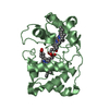
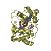
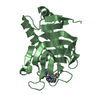
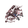
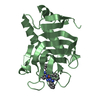
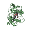
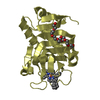
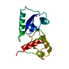
| Title | Crystal structure of oxidized recombinant cytochrome c4 from Pseudomonas stutzeri | ||||||
 Components Components | Cytochrome c4 | ||||||
 Keywords Keywords | ELECTRON TRANSPORT / diheme protein | ||||||
| Function / homology |  Function and homology information Function and homology informationelectron transfer activity / periplasmic space / iron ion binding / heme binding Similarity search - Function | ||||||
| Biological species |  Pseudomonas stutzeri (bacteria) Pseudomonas stutzeri (bacteria) | ||||||
| Method |  X-RAY DIFFRACTION / SYNCHROTRON / MOLECULAR REPLACEMENT / Resolution: 1.25 Å X-RAY DIFFRACTION / SYNCHROTRON / MOLECULAR REPLACEMENT / Resolution: 1.25 Å | ||||||
 Authors Authors | Noergaard, A. / Harris, P. / Larsen, S. / Christensen, H.E.M. | ||||||
 Citation Citation | Journal: To be Published Title: Structural comparison of recombinant Pseudomonas stutzeri cytochrome c4 in two oxidation states Authors: Noergaard, A. / Harris, P. / Larsen, S. / Christensen, H.E.M. #1: Journal: Acta Crystallogr.,Sect.D / Year: 1995Title: Crystallization and preliminary crystallographic investigations of cytochrome c4 from Pseudomonas stutzeri Authors: Kadziola, A. / Larsen, S. / Christensen, H.M. / Karlsson, J.-J. / Ulstrup, J. #2: Journal: Structure / Year: 1997Title: Crystal structure of the dihaem cytochrome c4 from Pseudomonas stutzeri determined at 2.2 A resolution Authors: Kadziola, A. / Larsen, S. #3: Journal: HANDBOOK OF METALLOPROTEINS / Year: 2001Title: Cytochrome c4 Authors: Andersen, N.H. / Christensen, H.E.M. / Iversen, G. / Noergaard, A. / Scharnagl, C. / Thuesen, M.H. / Ulstrup, J. | ||||||
| History |
|
- Structure visualization
| Structure viewer | Molecule: MolmilJmol/JSmol |
|---|
- Downloads & links
-Download
| PDBx/mmCIF format | 1m70.cif.gz | 335.4 KB | Display | PDBx/mmCIF format |
|---|---|---|---|---|
| PDB format | pdb1m70.ent.gz | 275.2 KB | Display | PDB format |
| PDBx/mmJSON format | 1m70.json.gz | Tree view | PDBx/mmJSON format | |
| Others |  Other downloads Other downloads |
-Validation report
| Arichive directory | https://data.pdbj.org/pub/pdb/validation_reports/m7/1m70ftp://data.pdbj.org/pub/pdb/validation_reports/m7/1m70 | HTTPS FTP |
|---|
-Related structure data
| Related structure data | 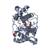 1m6zC 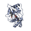 1etpS S: Starting model for refinement C: citing same article ( |
|---|---|
| Similar structure data |


-Links
 PDBj
PDBj


- Assembly
| Deposited unit | 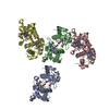
| ||||||||
|---|---|---|---|---|---|---|---|---|---|
| 1 | 
| ||||||||
| 2 | 
| ||||||||
| 3 | 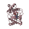
| ||||||||
| 4 | 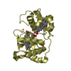
| ||||||||
| Unit cell |
|
-Components
| #1: Protein | Mass: 19700.062 Da / Num. of mol.: 4 Source method: isolated from a genetically manipulated source Source: (gene. exp.) Pseudomonas stutzeri (bacteria) / Plasmid: pNM185 / Production host: Pseudomonas putida (bacteria) / Strain (production host): PaW340 / References: UniProt: Q52369#2: Chemical | ChemComp-HEC /   Mass: 618.503 Da / Num. of mol.: 8 / Source method: obtained synthetically / Formula: C34H34FeN4O4 Mass: 618.503 Da / Num. of mol.: 8 / Source method: obtained synthetically / Formula: C34H34FeN4O4#3: Chemical | ChemComp-GOL / |   Mass: 92.094 Da / Num. of mol.: 1 / Source method: obtained synthetically / Formula: C3H8O3 Mass: 92.094 Da / Num. of mol.: 1 / Source method: obtained synthetically / Formula: C3H8O3#4: Water | ChemComp-HOH / |  Mass: 18.015 Da / Num. of mol.: 819 / Source method: isolated from a natural source / Formula: H2O Mass: 18.015 Da / Num. of mol.: 819 / Source method: isolated from a natural source / Formula: H2OHas protein modification | Y | |
|---|
-Experimental details
-Experiment
| Experiment | Method: X-RAY DIFFRACTION / Number of used crystals: 1 |
|---|
- Sample preparation
| Crystal | Density Matthews: 1.76 Å3/Da / Density % sol: 30.2 % |
|---|---|
| Crystal grow | Temperature: 298 K / Method: vapor diffusion, hanging drop / pH: 6.6 Details: 0.2M ammonium acetate, 0.1M sodium citrate pH 5.6, 30%(w/v) PEG 4000, pH 6.6, VAPOR DIFFUSION, HANGING DROP, temperature 298K |
-Data collection
| Diffraction |
| ||||||||||||||||||
|---|---|---|---|---|---|---|---|---|---|---|---|---|---|---|---|---|---|---|---|
| Diffraction source |
| ||||||||||||||||||
| Detector |
| ||||||||||||||||||
| Radiation |
| ||||||||||||||||||
| Radiation wavelength |
| ||||||||||||||||||
| Reflection | Resolution: 1.25→30 Å / Num. all: 178812 / Num. obs: 175219 / % possible obs: 90.9 % / Observed criterion σ(F): 0 / Observed criterion σ(I): 0 | ||||||||||||||||||
| Reflection shell | Resolution: 1.25→1.29 Å / % possible all: 77 |

- Processing
| Software |
| |||||||||||||||||||||||||||||||||
|---|---|---|---|---|---|---|---|---|---|---|---|---|---|---|---|---|---|---|---|---|---|---|---|---|---|---|---|---|---|---|---|---|---|---|
| Refinement | Method to determine structure: MOLECULAR REPLACEMENT Starting model: PDB entry 1ETP Resolution: 1.25→30 Å / Num. parameters: 60529 / Num. restraintsaints: 86801 / Cross valid method: THROUGHOUT / σ(F): 0 / Stereochemistry target values: Engh & Huber / Details: Anisotropic refinement. Riding H-atoms introduced.
| |||||||||||||||||||||||||||||||||
| Solvent computation | Solvent model: MOEWS & KRETSINGER, J.MOL.BIOL.91(1973)201-228 | |||||||||||||||||||||||||||||||||
| Refine analyze | Num. disordered residues: 10 / Occupancy sum hydrogen: 5368 / Occupancy sum non hydrogen: 6688 | |||||||||||||||||||||||||||||||||
| Refinement step | Cycle: LAST / Resolution: 1.25→30 Å
| |||||||||||||||||||||||||||||||||
| Refine LS restraints |
|
