 Movie
Movie Controller


+ Open data

- Basic information
| Entry | Database: PDB / ID: 1etp | ||||||
|---|---|---|---|---|---|---|---|
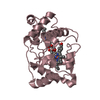
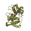
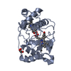
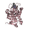
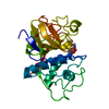
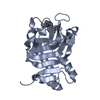
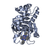
| Title | CRYSTAL STRUCTURE OF CYTOCHROME C4 FROM PSEUDOMONAS STUTZERI | ||||||
 Components Components | CYTOCHROME C4 | ||||||
 Keywords Keywords | ELECTRON TRANSPORT / CYTOCHROME C4 / DIHEME PROTEIN / PSEUDOMONAS STUTZERI | ||||||
| Function / homology |  Function and homology information Function and homology informationperiplasmic space / electron transfer activity / iron ion binding / heme binding Similarity search - Function | ||||||
| Biological species |  Pseudomonas stutzeri (bacteria) Pseudomonas stutzeri (bacteria) | ||||||
| Method |  X-RAY DIFFRACTION / Resolution: 2.2 Å X-RAY DIFFRACTION / Resolution: 2.2 Å | ||||||
 Authors Authors | Kadziola, A. / Larsen, S. | ||||||
 Citation Citation | Journal: Structure / Year: 1997 Title: Crystal structure of the dihaem cytochrome c4 from Pseudomonas stutzeri determined at 2.2A resolution. Authors: Kadziola, A. / Larsen, S. #1: Journal: Eur.J.Biochem. / Year: 1995Title: Electron Transfer and Spectral Alpha-Band Properties of the Di-Heme Protein Cytochrome C4 from Pseudomonas Stutzeri Authors: Conrad, L.S. / Karlsson, J.J. / Ulstrup, J. #2: Journal: Acta Crystallogr.,Sect.D / Year: 1995Title: Crystallization and Preliminary Crystallographic Investigations of Cytochrome C4 from Pseudomonas Stutzeri Authors: Kadziola, A. / Larsen, S. / Christensen, H.M. / Karlsson, J.-J. / Ulstrup, J. #3: Journal: Gene / Year: 1994Title: Cloning and Characterisation of the Gene Encoding Cytochrome C4 from Pseudomonas Stutzeri Authors: Christensen, H.E. | ||||||
| History |
|
- Structure visualization
| Structure viewer | Molecule: MolmilJmol/JSmol |
|---|
- Downloads & links
-Download
| PDBx/mmCIF format | 1etp.cif.gz | 87.6 KB | Display | PDBx/mmCIF format |
|---|---|---|---|---|
| PDB format | pdb1etp.ent.gz | 67.2 KB | Display | PDB format |
| PDBx/mmJSON format | 1etp.json.gz | Tree view | PDBx/mmJSON format | |
| Others |  Other downloads Other downloads |
-Validation report
| Arichive directory | https://data.pdbj.org/pub/pdb/validation_reports/et/1etpftp://data.pdbj.org/pub/pdb/validation_reports/et/1etp | HTTPS FTP |
|---|
-Related structure data
| Similar structure data |
|---|


-Links
 PDBj
PDBj


- Assembly
| Deposited unit | 
| ||||||||
|---|---|---|---|---|---|---|---|---|---|
| 1 | 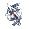
| ||||||||
| 2 | 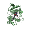
| ||||||||
| Unit cell |
| ||||||||
| Noncrystallographic symmetry (NCS) | NCS oper: (Code: given Matrix: (-0.798002, 0.444533, -0.40692), Vector: |
-Components
| #1: Protein | Mass: 19700.062 Da / Num. of mol.: 2 / Source method: isolated from a natural source / Source: (natural) Pseudomonas stutzeri (bacteria) / References: UniProt: Q52369#2: Chemical | ChemComp-HEM /   Mass: 616.487 Da / Num. of mol.: 4 / Source method: obtained synthetically / Formula: C34H32FeN4O4 Mass: 616.487 Da / Num. of mol.: 4 / Source method: obtained synthetically / Formula: C34H32FeN4O4#3: Water | ChemComp-HOH / |  Mass: 18.015 Da / Num. of mol.: 138 / Source method: isolated from a natural source / Formula: H2O Mass: 18.015 Da / Num. of mol.: 138 / Source method: isolated from a natural source / Formula: H2OCompound details | RESIDUES HIS 18 AND MET 66 FORM HEME IRON LIGAND BONDS TO THE N-TERMINAL HEME WHILE HIS 123 AND MET ...RESIDUES HIS 18 AND MET 66 FORM HEME IRON LIGAND BONDS TO THE N-TERMINAL HEME WHILE HIS 123 AND MET 167 FORM HEME IRON LIGAND BONDS TO THE C-TERMINAL HEME. | Has protein modification | Y | Nonpolymer details | THE DEPOSITOR INDICATED THAT THE CHARGE ON THE HEME GROUP IS 2+ OR 3+. | |
|---|
-Experimental details
-Experiment
| Experiment | Method: X-RAY DIFFRACTION |
|---|
- Sample preparation
| Crystal | Density Matthews: 2.32 Å3/Da / Density % sol: 39 % | ||||||||||||||||||||||||||||||||||||||||||||||||||||||||||||
|---|---|---|---|---|---|---|---|---|---|---|---|---|---|---|---|---|---|---|---|---|---|---|---|---|---|---|---|---|---|---|---|---|---|---|---|---|---|---|---|---|---|---|---|---|---|---|---|---|---|---|---|---|---|---|---|---|---|---|---|---|---|
| Crystal grow | pH: 5.6 / Details: pH 5.6 | ||||||||||||||||||||||||||||||||||||||||||||||||||||||||||||
| Crystal grow | *PLUS Temperature: 293 K / pH: 7.5 / Method: vapor diffusion, hanging dropDetails: Kadziola, A., (1995) Acta Crystallogr.,Sect.D, 51, 1071. | ||||||||||||||||||||||||||||||||||||||||||||||||||||||||||||
| Components of the solutions | *PLUS
|
-Data collection
| Diffraction | Mean temperature: 274 K |
|---|---|
| Diffraction source | Source: ROTATING ANODE / Wavelength: 1.5418 |
| Detector | Type: RIGAKU / Detector: IMAGE PLATE / Date: Mar 12, 1993 |
| Radiation | Monochromator: GRAPHITE(002) / Monochromatic (M) / Laue (L): M / Scattering type: x-ray |
| Radiation wavelength | Wavelength: 1.5418 Å / Relative weight: 1 |
| Reflection | Resolution: 2.2→58.7 Å / Num. obs: 18020 / % possible obs: 98.1 % / Observed criterion σ(I): 0 / Redundancy: 3.8 % / Rsym value: 0.033 / Net I/σ(I): 13.5 |
| Reflection | *PLUS Rmerge(I) obs: 0.033 |
- Processing
| Software |
| ||||||||||||||||||||||||||||||||||||||||||||||||||||||||||||
|---|---|---|---|---|---|---|---|---|---|---|---|---|---|---|---|---|---|---|---|---|---|---|---|---|---|---|---|---|---|---|---|---|---|---|---|---|---|---|---|---|---|---|---|---|---|---|---|---|---|---|---|---|---|---|---|---|---|---|---|---|---|
| Refinement | Resolution: 2.2→8 Å / σ(F): 0 Details: RAMACHANDRAN PLOT ONE RESIDUE IN EACH MOLECULE (LISTED BELOW) ARE FOUND IN THE 'FORBIDDEN' REGION OF THE RAMACHANDRAN PLOT. BOTH RESIDUES ARE SITUATED IN REGIONS WITH WELL DEFINED ELECTRON ...Details: RAMACHANDRAN PLOT ONE RESIDUE IN EACH MOLECULE (LISTED BELOW) ARE FOUND IN THE 'FORBIDDEN' REGION OF THE RAMACHANDRAN PLOT. BOTH RESIDUES ARE SITUATED IN REGIONS WITH WELL DEFINED ELECTRON DENSITY. PHI PSI OMEGA 1 ALA A 124 65.95 149.82 -180.00 2 ALA B 124 76.18 151.83 -179.44
| ||||||||||||||||||||||||||||||||||||||||||||||||||||||||||||
| Displacement parameters | Biso mean: 22.8 Å2 | ||||||||||||||||||||||||||||||||||||||||||||||||||||||||||||
| Refine analyze | Luzzati coordinate error obs: 0.35 Å | ||||||||||||||||||||||||||||||||||||||||||||||||||||||||||||
| Refinement step | Cycle: LAST / Resolution: 2.2→8 Å
| ||||||||||||||||||||||||||||||||||||||||||||||||||||||||||||
| Refine LS restraints |
| ||||||||||||||||||||||||||||||||||||||||||||||||||||||||||||
| Software | *PLUS Name: X-PLOR / Version: 3.1 / Classification: refinement | ||||||||||||||||||||||||||||||||||||||||||||||||||||||||||||
| Refine LS restraints | *PLUS
|
