 Movie
Movie Controller

[English] 日本語
 Yorodumi
- PDB-3h8b: A combined crystallographic and molecular dynamics study of cathe... -
+ Open data

- Basic information
| Entry | Database: PDB / ID: 3h8b | ||||||
|---|---|---|---|---|---|---|---|
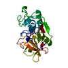
| Title | A combined crystallographic and molecular dynamics study of cathepsin-L retro-binding inhibitors(compound 9) | ||||||
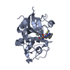
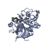
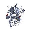
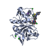
 Components Components | Cathepsin L1 | ||||||
 Keywords Keywords | HYDROLASE / cysteine proteases / Cathepsin L / Disulfide bond / Glycoprotein / Lysosome / Protease / Thiol protease / Zymogen | ||||||
| Function / homology |  Function and homology information Function and homology informationenkephalin processing / cathepsin L / CD4-positive, alpha-beta T cell lineage commitment / macrophage apoptotic process / chromaffin granule / antigen processing and presentation of peptide antigen / elastin catabolic process / HS-GAG degradation / RUNX1 regulates transcription of genes involved in differentiation of keratinocytes / endolysosome lumen ...enkephalin processing / cathepsin L / CD4-positive, alpha-beta T cell lineage commitment / macrophage apoptotic process / chromaffin granule / antigen processing and presentation of peptide antigen / elastin catabolic process / HS-GAG degradation / RUNX1 regulates transcription of genes involved in differentiation of keratinocytes / endolysosome lumen / cellular response to thyroid hormone stimulus / Trafficking and processing of endosomal TLR / proteoglycan binding / Assembly of collagen fibrils and other multimeric structures / zymogen activation / antigen processing and presentation / Collagen degradation / protein autoprocessing / collagen catabolic process / fibronectin binding / serpin family protein binding / Degradation of the extracellular matrix / collagen binding / receptor-mediated endocytosis of virus by host cell / Attachment and Entry / multivesicular body / endocytic vesicle lumen / cysteine-type peptidase activity / MHC class II antigen presentation / lysosomal lumen / : / Endosomal/Vacuolar pathway / Degradation of CDH1 / antigen processing and presentation of exogenous peptide antigen via MHC class II / extracellular matrix / histone binding / adaptive immune response / Attachment and Entry / lysosome / apical plasma membrane / fusion of virus membrane with host plasma membrane / cysteine-type endopeptidase activity / fusion of virus membrane with host endosome membrane / symbiont entry into host cell / Golgi apparatus / proteolysis / : / extracellular exosome / extracellular region / nucleus / plasma membrane Similarity search - Function | ||||||
| Biological species |  Homo sapiens (human) Homo sapiens (human) | ||||||
| Method |  X-RAY DIFFRACTION / SYNCHROTRON / MOLECULAR REPLACEMENT / Resolution: 1.8 Å X-RAY DIFFRACTION / SYNCHROTRON / MOLECULAR REPLACEMENT / Resolution: 1.8 Å | ||||||
 Authors Authors | Tulsidas, S.R. / Chowdhury, S.F. / Kumar, S. / Joseph, L. / Purisima, E.O. / Sivaraman, J. | ||||||
 Citation Citation | Journal: J.Med.Chem. / Year: 2009 Title: A combined crystallographic and molecular dynamics study of cathepsin L retrobinding inhibitors Authors: Shenoy, R.T. / Chowdhury, S.F. / Kumar, S. / Joseph, L. / Purisima, E.O. / Sivaraman, J. | ||||||
| History |
|
- Structure visualization
| Structure viewer | Molecule: MolmilJmol/JSmol |
|---|
- Downloads & links
-Download
| PDBx/mmCIF format | 3h8b.cif.gz | 274.2 KB | Display | PDBx/mmCIF format |
|---|---|---|---|---|
| PDB format | pdb3h8b.ent.gz | 224.2 KB | Display | PDB format |
| PDBx/mmJSON format | 3h8b.json.gz | Tree view | PDBx/mmJSON format | |
| Others |  Other downloads Other downloads |
-Validation report
| Arichive directory | https://data.pdbj.org/pub/pdb/validation_reports/h8/3h8bftp://data.pdbj.org/pub/pdb/validation_reports/h8/3h8b | HTTPS FTP |
|---|
-Related structure data


-Links
 PDBj
PDBj


- Assembly
| Deposited unit | 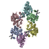
| ||||||||
|---|---|---|---|---|---|---|---|---|---|
| 1 | 
| ||||||||
| 2 | 
| ||||||||
| 3 | 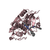
| ||||||||
| 4 | 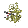
| ||||||||
| 5 | 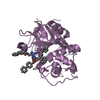
| ||||||||
| 6 | 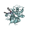
| ||||||||
| Unit cell |
|
-Components
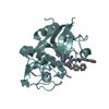
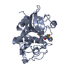
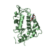
| #1: Protein | Mass: 24191.701 Da / Num. of mol.: 6 Fragment: Cathepsin L Heavy Chain and Light Chain, UNP residues 114-333 Source method: isolated from a genetically manipulated source Source: (gene. exp.) Homo sapiens (human) / Gene: Cathepsin L / Production host:  #2: Chemical | ChemComp-NSY / 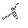  Mass: 941.169 Da / Num. of mol.: 6 / Source method: obtained synthetically / Formula: C57H64N8O5 Mass: 941.169 Da / Num. of mol.: 6 / Source method: obtained synthetically / Formula: C57H64N8O5#3: Water | ChemComp-HOH / |  Mass: 18.015 Da / Num. of mol.: 562 / Source method: isolated from a natural source / Formula: H2O Mass: 18.015 Da / Num. of mol.: 562 / Source method: isolated from a natural source / Formula: H2OHas protein modification | Y | |
|---|
-Experimental details
-Experiment
| Experiment | Method: X-RAY DIFFRACTION / Number of used crystals: 1 |
|---|
- Sample preparation
| Crystal | Density Matthews: 2.19 Å3/Da / Density % sol: 43.77 % |
|---|---|
| Crystal grow | Method: vapor diffusion, hanging drop / Details: VAPOR DIFFUSION, HANGING DROP |
-Data collection
| Diffraction | Mean temperature: 100 K |
|---|---|
| Diffraction source | Source: SYNCHROTRON / Site: NSLS  / Beamline: X29A / Wavelength: 1 Å / Beamline: X29A / Wavelength: 1 Å |
| Detector | Type: ADSC QUANTUM 315 / Detector: CCD |
| Radiation | Protocol: SINGLE WAVELENGTH / Monochromatic (M) / Laue (L): M / Scattering type: x-ray |
| Radiation wavelength | Wavelength: 1 Å / Relative weight: 1 |
| Reflection | Resolution: 1.79→50 Å / Num. obs: 116974 / Rsym value: 0.06 / Net I/σ(I): 17.1 |
- Processing
| Software |
| ||||||||||||||||||||
|---|---|---|---|---|---|---|---|---|---|---|---|---|---|---|---|---|---|---|---|---|---|
| Refinement | Method to determine structure: MOLECULAR REPLACEMENT / Resolution: 1.8→45 Å / Occupancy max: 1 / Occupancy min: 1 / Cross valid method: THROUGHOUT / σ(F): 3874
| ||||||||||||||||||||
| Solvent computation | Bsol: 40.919 Å2 | ||||||||||||||||||||
| Displacement parameters | Biso max: 101.66 Å2 / Biso mean: 27.671 Å2 / Biso min: 8.22 Å2
| ||||||||||||||||||||
| Refinement step | Cycle: LAST / Resolution: 1.8→45 Å
| ||||||||||||||||||||
| Refine LS restraints |
|
