[English] 日本語
 Yorodumi
- PDB-7aft: Cryo-EM structure of the signal sequence-engaged post-translation... -
+ Open data
- Basic information
| Entry | Database: PDB / ID: 7aft | |||||||||||||||||||||||||||||||||||||||||||||
|---|---|---|---|---|---|---|---|---|---|---|---|---|---|---|---|---|---|---|---|---|---|---|---|---|---|---|---|---|---|---|---|---|---|---|---|---|---|---|---|---|---|---|---|---|---|---|
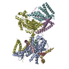
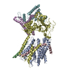
| Title | Cryo-EM structure of the signal sequence-engaged post-translational Sec translocon | |||||||||||||||||||||||||||||||||||||||||||||
 Components Components |
| |||||||||||||||||||||||||||||||||||||||||||||
 Keywords Keywords | MEMBRANE PROTEIN / Post-translational translocation / Protein translocation / Sec complex / Signal sequence | |||||||||||||||||||||||||||||||||||||||||||||
| Function / homology |  Function and homology information Function and homology informationmating pheromone activity / mating / misfolded protein transport / Sec62/Sec63 complex / translocon complex / Insertion of tail-anchored proteins into the endoplasmic reticulum membrane / pheromone-dependent signal transduction involved in conjugation with cellular fusion / rough endoplasmic reticulum membrane / cytosol to endoplasmic reticulum transport / Ssh1 translocon complex ...mating pheromone activity / mating / misfolded protein transport / Sec62/Sec63 complex / translocon complex / Insertion of tail-anchored proteins into the endoplasmic reticulum membrane / pheromone-dependent signal transduction involved in conjugation with cellular fusion / rough endoplasmic reticulum membrane / cytosol to endoplasmic reticulum transport / Ssh1 translocon complex / Sec61 translocon complex / protein-transporting ATPase activity / post-translational protein targeting to endoplasmic reticulum membrane / SRP-dependent cotranslational protein targeting to membrane, translocation / filamentous growth / signal sequence binding / SRP-dependent cotranslational protein targeting to membrane / post-translational protein targeting to membrane, translocation / cupric ion binding / peptide transmembrane transporter activity / nuclear inner membrane / retrograde protein transport, ER to cytosol / protein transmembrane transporter activity / ERAD pathway / guanyl-nucleotide exchange factor activity / cell periphery / ribosome binding / endoplasmic reticulum membrane / structural molecule activity / endoplasmic reticulum / mitochondrion / extracellular region / membrane / cytosol Similarity search - Function | |||||||||||||||||||||||||||||||||||||||||||||
| Biological species |  | |||||||||||||||||||||||||||||||||||||||||||||
| Method | ELECTRON MICROSCOPY / single particle reconstruction / cryo EM / Resolution: 4.4 Å | |||||||||||||||||||||||||||||||||||||||||||||
 Authors Authors | Weng, T.-H. / Beatrix, B. / Berninghausen, O. / Becker, T. / Cheng, J. / Beckmann, R. | |||||||||||||||||||||||||||||||||||||||||||||
 Citation Citation |  Journal: EMBO J / Year: 2021 Journal: EMBO J / Year: 2021Title: Architecture of the active post-translational Sec translocon. Authors: Tsai-Hsuan Weng / Wieland Steinchen / Birgitta Beatrix / Otto Berninghausen / Thomas Becker / Gert Bange / Jingdong Cheng / Roland Beckmann /  Abstract: In eukaryotes, most secretory and membrane proteins are targeted by an N-terminal signal sequence to the endoplasmic reticulum, where the trimeric Sec61 complex serves as protein-conducting channel ...In eukaryotes, most secretory and membrane proteins are targeted by an N-terminal signal sequence to the endoplasmic reticulum, where the trimeric Sec61 complex serves as protein-conducting channel (PCC). In the post-translational mode, fully synthesized proteins are recognized by a specialized channel additionally containing the Sec62, Sec63, Sec71, and Sec72 subunits. Recent structures of this Sec complex in the idle state revealed the overall architecture in a pre-opened state. Here, we present a cryo-EM structure of the yeast Sec complex bound to a substrate, and a crystal structure of the Sec62 cytosolic domain. The signal sequence is inserted into the lateral gate of Sec61α similar to previous structures, yet, with the gate adopting an even more open conformation. The signal sequence is flanked by two Sec62 transmembrane helices, the cytoplasmic N-terminal domain of Sec62 is more rigidly positioned, and the plug domain is relocated. We crystallized the Sec62 domain and mapped its interaction with the C-terminus of Sec63. Together, we obtained a near-complete and integrated model of the active Sec complex. | |||||||||||||||||||||||||||||||||||||||||||||
| History |
|
- Structure visualization
| Movie |
 Movie viewer Movie viewer |
|---|---|
| Structure viewer | Molecule:  Molmil Molmil Jmol/JSmol Jmol/JSmol |
- Downloads & links
- Download
| PDBx/mmCIF format |  7aft.cif.gz 7aft.cif.gz | 218.9 KB | Display |  PDBx/mmCIF format PDBx/mmCIF format |
|---|---|---|---|---|
| PDB format |  pdb7aft.ent.gz pdb7aft.ent.gz | 139.7 KB | Display |  PDB format PDB format |
| PDBx/mmJSON format |  7aft.json.gz 7aft.json.gz | Tree view |  PDBx/mmJSON format PDBx/mmJSON format | |
| Others |  Other downloads Other downloads |
-Validation report
| Summary document |  7aft_validation.pdf.gz 7aft_validation.pdf.gz | 933 KB | Display |  wwPDB validaton report wwPDB validaton report |
|---|---|---|---|---|
| Full document |  7aft_full_validation.pdf.gz 7aft_full_validation.pdf.gz | 936.1 KB | Display | |
| Data in XML |  7aft_validation.xml.gz 7aft_validation.xml.gz | 35 KB | Display | |
| Data in CIF |  7aft_validation.cif.gz 7aft_validation.cif.gz | 58.5 KB | Display | |
| Arichive directory |  https://data.pdbj.org/pub/pdb/validation_reports/af/7aft https://data.pdbj.org/pub/pdb/validation_reports/af/7aft ftp://data.pdbj.org/pub/pdb/validation_reports/af/7aft ftp://data.pdbj.org/pub/pdb/validation_reports/af/7aft | HTTPS FTP |
-Related structure data
| Related structure data |  11774MC  6zzzC M: map data used to model this data C: citing same article ( |
|---|---|
| Similar structure data |
- Links
Links
- Assembly
| Deposited unit | 
|
|---|---|
| 1 |
|
- Components
-Protein transport protein ... , 3 types, 3 molecules ABC
| #1: Protein | Mass: 52978.148 Da / Num. of mol.: 1 / Source method: isolated from a natural source Source: (natural)  Strain: ATCC 204508 / S288c / References: UniProt: P32915 |
|---|---|
| #2: Protein | Mass: 8723.155 Da / Num. of mol.: 1 / Source method: isolated from a natural source Source: (natural)  Strain: ATCC 204508 / S288c / References: UniProt: P52870 |
| #3: Protein | Mass: 8958.641 Da / Num. of mol.: 1 / Source method: isolated from a natural source Source: (natural)  Strain: ATCC 204508 / S288c / References: UniProt: P35179 |
-Protein , 2 types, 2 molecules DH
| #4: Protein | Mass: 75432.258 Da / Num. of mol.: 1 / Source method: isolated from a natural source Source: (natural)  Strain: ATCC 204508 / S288c / References: UniProt: P14906 |
|---|---|
| #8: Protein | Mass: 19766.402 Da / Num. of mol.: 1 Source method: isolated from a genetically manipulated source Source: (gene. exp.)  Strain: ATCC 204508 / S288c / Gene: MF(ALPHA)1, MF-ALPHA-1, MFAL1, YPL187W / Plasmid: pET28a / Production host:  |
-Translocation protein ... , 3 types, 3 molecules EFG
| #5: Protein | Mass: 24263.939 Da / Num. of mol.: 1 / Source method: isolated from a natural source Source: (natural)  Strain: ATCC 204508 / S288c / References: UniProt: P33754 |
|---|---|
| #6: Protein | Mass: 21631.090 Da / Num. of mol.: 1 / Source method: isolated from a natural source Source: (natural)  Strain: ATCC 204508 / S288c / References: UniProt: P39742 |
| #7: Protein | Mass: 35649.891 Da / Num. of mol.: 1 Source method: isolated from a genetically manipulated source Details: The residue numbering in chain G is based on the transmembrane helix prediction of Sec62. Source: (gene. exp.)  Strain: ATCC 204508 / S288c / Gene: SEC62, YPL094C, LPG14C / Production host:  |
-Details
| Has protein modification | N |
|---|
-Experimental details
-Experiment
| Experiment | Method: ELECTRON MICROSCOPY |
|---|---|
| EM experiment | Aggregation state: PARTICLE / 3D reconstruction method: single particle reconstruction |
- Sample preparation
| Component |
| ||||||||||||||||||||||||||||||
|---|---|---|---|---|---|---|---|---|---|---|---|---|---|---|---|---|---|---|---|---|---|---|---|---|---|---|---|---|---|---|---|
| Molecular weight | Value: 0.255 MDa / Experimental value: NO | ||||||||||||||||||||||||||||||
| Source (natural) |
| ||||||||||||||||||||||||||||||
| Source (recombinant) |
| ||||||||||||||||||||||||||||||
| Buffer solution | pH: 7.4 | ||||||||||||||||||||||||||||||
| Buffer component |
| ||||||||||||||||||||||||||||||
| Specimen | Conc.: 5 mg/ml / Embedding applied: NO / Shadowing applied: NO / Staining applied: NO / Vitrification applied: YES | ||||||||||||||||||||||||||||||
| Specimen support | Grid material: GOLD / Grid type: UltrAuFoil R2/2 | ||||||||||||||||||||||||||||||
| Vitrification | Instrument: FEI VITROBOT MARK IV / Cryogen name: ETHANE / Humidity: 100 % / Chamber temperature: 277 K |
- Electron microscopy imaging
| Experimental equipment |  Model: Titan Krios / Image courtesy: FEI Company | ||||||||||||||||||
|---|---|---|---|---|---|---|---|---|---|---|---|---|---|---|---|---|---|---|---|
| Microscopy | Model: FEI TITAN KRIOS | ||||||||||||||||||
| Electron gun | Electron source:  FIELD EMISSION GUN / Accelerating voltage: 300 kV / Illumination mode: FLOOD BEAM FIELD EMISSION GUN / Accelerating voltage: 300 kV / Illumination mode: FLOOD BEAM | ||||||||||||||||||
| Electron lens | Mode: BRIGHT FIELD | ||||||||||||||||||
| Image recording |
| ||||||||||||||||||
| Image scans | Movie frames/image: 40 |
- Processing
| Software | Name: PHENIX / Version: 1.17.1_3660: / Classification: refinement | ||||||||||||||||||||||||||||||||||||||||||||||||||||||||||||||||||||||
|---|---|---|---|---|---|---|---|---|---|---|---|---|---|---|---|---|---|---|---|---|---|---|---|---|---|---|---|---|---|---|---|---|---|---|---|---|---|---|---|---|---|---|---|---|---|---|---|---|---|---|---|---|---|---|---|---|---|---|---|---|---|---|---|---|---|---|---|---|---|---|---|
| EM software |
| ||||||||||||||||||||||||||||||||||||||||||||||||||||||||||||||||||||||
| Image processing |
| ||||||||||||||||||||||||||||||||||||||||||||||||||||||||||||||||||||||
| CTF correction |
| ||||||||||||||||||||||||||||||||||||||||||||||||||||||||||||||||||||||
| 3D reconstruction | Algorithm: FOURIER SPACE / Entry-ID: 7AFT / Resolution: 4.4 Å / Resolution method: FSC 0.143 CUT-OFF / Symmetry type: POINT
| ||||||||||||||||||||||||||||||||||||||||||||||||||||||||||||||||||||||
| Atomic model building | Protocol: RIGID BODY FIT | ||||||||||||||||||||||||||||||||||||||||||||||||||||||||||||||||||||||
| Atomic model building | PDB-ID: 6N3Q Accession code: 6N3Q / Source name: PDB / Type: experimental model | ||||||||||||||||||||||||||||||||||||||||||||||||||||||||||||||||||||||
| Refine LS restraints |
|
 Movie
Movie Controller


 PDBj
PDBj


