Movie
Movie Controller


+ Open data

- Basic information
| Entry | Database: PDB / ID: 6zxa | ||||||||||||
|---|---|---|---|---|---|---|---|---|---|---|---|---|---|
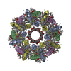
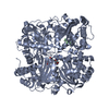
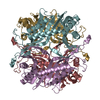
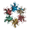
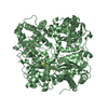
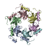
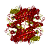
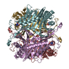
| Title | LH2 complex from Marichromatium purpuratum | ||||||||||||
 Components Components |
| ||||||||||||
 Keywords Keywords | PHOTOSYNTHESIS / purple bacteria / light harvesting complex / Marichromatium purpuratum / okenone. | ||||||||||||
| Function / homology |  Function and homology information Function and homology informationlight-harvesting complex / plasma membrane light-harvesting complex / bacteriochlorophyll binding / photosynthesis, light reaction / metal ion binding / plasma membrane Similarity search - Function | ||||||||||||
| Biological species |  Marichromatium purpuratum 984 (bacteria) Marichromatium purpuratum 984 (bacteria) | ||||||||||||
| Method | ELECTRON MICROSCOPY / single particle reconstruction / cryo EM / Resolution: 2.38 Å | ||||||||||||
 Authors Authors | Gardiner, A.T. / Naydenova, K. / Castro-Hartmann, P. / Nguyen-Phan, T.C. / Russo, C.J. / Sader, K. / Hunter, C.N. / Cogdell, R.J. / Qian, P. | ||||||||||||
| Funding support |  United States, United States,  United Kingdom, 3items United Kingdom, 3items
| ||||||||||||
 Citation Citation | Journal: Sci Adv / Year: 2021 Title: The 2.4 Å cryo-EM structure of a heptameric light-harvesting 2 complex reveals two carotenoid energy transfer pathways. Authors: Alastair T Gardiner / Katerina Naydenova / Pablo Castro-Hartmann / Tu C Nguyen-Phan / Christopher J Russo / Kasim Sader / C Neil Hunter / Richard J Cogdell / Pu Qian /  Abstract: We report the 2.4 Ångström resolution structure of the light-harvesting 2 (LH2) complex from () determined by cryogenic electron microscopy. The structure contains a heptameric ring that is ...We report the 2.4 Ångström resolution structure of the light-harvesting 2 (LH2) complex from () determined by cryogenic electron microscopy. The structure contains a heptameric ring that is unique among all known LH2 structures, explaining the unusual spectroscopic properties of this bacterial antenna complex. We identify two sets of distinct carotenoids in the structure and describe a network of energy transfer pathways from the carotenoids to bacteriochlorophyll molecules. The geometry imposed by the heptameric ring controls the resonant coupling of the long-wavelength energy absorption band. Together, these details reveal key aspects of the assembly and oligomeric form of purple bacterial LH2 complexes that were previously inaccessible by any technique. | ||||||||||||
| History |
|
- Structure visualization
| Movie |
Movie viewer |
|---|---|
| Structure viewer | Molecule: MolmilJmol/JSmol |
- Downloads & links
-Download
| PDBx/mmCIF format | 6zxa.cif.gz | 248 KB | Display | PDBx/mmCIF format |
|---|---|---|---|---|
| PDB format | pdb6zxa.ent.gz | 183.7 KB | Display | PDB format |
| PDBx/mmJSON format | 6zxa.json.gz | Tree view | PDBx/mmJSON format | |
| Others |  Other downloads Other downloads |
-Validation report
| Arichive directory | https://data.pdbj.org/pub/pdb/validation_reports/zx/6zxaftp://data.pdbj.org/pub/pdb/validation_reports/zx/6zxa | HTTPS FTP |
|---|
-Related structure data
| Related structure data |  11516MC M: map data used to model this data C: citing same article ( |
|---|---|
| Similar structure data | |
| EM raw data | EMPIAR-10606 (Title: Movies of a heptameric light-harvesting 2 complex from the marine purple bacterium Mch. purpuratum Data size: 12.2 TB Data #1: Unaligned multi-frame micrographs of purple bacterial LH2 on HexAuFoil supports [micrographs - multiframe]) |
-Links
 PDBj
PDBj- Assembly
| Deposited unit | 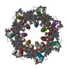
|
|---|---|
| 1 |
|
-Components
| #1: Protein | Mass: 8015.369 Da / Num. of mol.: 7 / Source method: isolated from a natural source / Source: (natural) Marichromatium purpuratum 984 (bacteria) / References: UniProt: W0E6A1#2: Protein/peptide | Mass: 5554.275 Da / Num. of mol.: 7 / Source method: isolated from a natural source / Source: (natural) Marichromatium purpuratum 984 (bacteria) / References: UniProt: W0E5B0#3: Chemical | ChemComp-BCL /   Mass: 911.504 Da / Num. of mol.: 21 / Source method: obtained synthetically / Formula: C55H74MgN4O6 / Feature type: SUBJECT OF INVESTIGATION Mass: 911.504 Da / Num. of mol.: 21 / Source method: obtained synthetically / Formula: C55H74MgN4O6 / Feature type: SUBJECT OF INVESTIGATION#4: Chemical | ChemComp-QS2 / 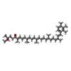  Mass: 578.866 Da / Num. of mol.: 7 / Source method: obtained synthetically / Formula: C41H54O2 / Feature type: SUBJECT OF INVESTIGATION Mass: 578.866 Da / Num. of mol.: 7 / Source method: obtained synthetically / Formula: C41H54O2 / Feature type: SUBJECT OF INVESTIGATION#5: Chemical | ChemComp-QSE /   Mass: 578.866 Da / Num. of mol.: 7 / Source method: obtained synthetically / Formula: C41H54O2 / Feature type: SUBJECT OF INVESTIGATION Mass: 578.866 Da / Num. of mol.: 7 / Source method: obtained synthetically / Formula: C41H54O2 / Feature type: SUBJECT OF INVESTIGATIONHas ligand of interest | Y | |
|---|
-Experimental details
-Experiment
| Experiment | Method: ELECTRON MICROSCOPY |
|---|---|
| EM experiment | Aggregation state: PARTICLE / 3D reconstruction method: single particle reconstruction |
- Sample preparation
| Component | Name: Light harvesting complex 2 (LH2) / Type: COMPLEX Details: The LH2 complex from Mar. purpuratum forms a circular heptameter. Each subunit consists of an alpha/beta pair, in which all pigment molecules, three Bchl a and two carotenoids are bound non-covalently. Entity ID: #1-#2 / Source: NATURAL |
|---|---|
| Molecular weight | Value: 0.11 MDa / Experimental value: NO |
| Source (natural) | Organism: Marichromatium purpuratum (bacteria) / Strain: BN5500 |
| Buffer solution | pH: 8 Details: The final buffer contains 0.02 % n-dodecyl-beta-D-maltopyranoside (DDM) |
| Buffer component | Conc.: 20 mM/L / Name: Tris.HCl |
| Specimen | Conc.: 5.5 mg/ml / Embedding applied: NO / Shadowing applied: NO / Staining applied: NO / Vitrification applied: YES Details: The sample was mono disperse with detergent beta-DDM. |
| Specimen support | Details: Fischione 1070 / Grid material: GOLD / Grid mesh size: 400 divisions/in. / Grid type: Homemade |
| Vitrification | Instrument: HOMEMADE PLUNGER / Cryogen name: ETHANE / Humidity: 100 % / Chamber temperature: 277 K / Details: Blowing time, 10.0 sec. |
- Electron microscopy imaging
| Experimental equipment |  Model: Titan Krios / Image courtesy: FEI Company |
|---|---|
| Microscopy | Model: FEI TITAN KRIOS |
| Electron gun | Electron source:  FIELD EMISSION GUN / Accelerating voltage: 300 kV / Illumination mode: FLOOD BEAM FIELD EMISSION GUN / Accelerating voltage: 300 kV / Illumination mode: FLOOD BEAM |
| Electron lens | Mode: BRIGHT FIELD / Nominal magnification: 120000 X / Nominal defocus max: 2400 nm / Nominal defocus min: 800 nm / Calibrated defocus min: 400 nm / Calibrated defocus max: 3000 nm / Cs: 2.7 mm / C2 aperture diameter: 50 µm / Alignment procedure: COMA FREE |
| Specimen holder | Cryogen: NITROGEN / Specimen holder model: FEI TITAN KRIOS AUTOGRID HOLDER / Temperature (min): 80 K |
| Image recording | Average exposure time: 0.29 sec. / Electron dose: 43.7 e/Å2 / Film or detector model: FEI FALCON IV (4k x 4k) / Num. of grids imaged: 1 / Num. of real images: 9543 |
- Processing
| Software |
| ||||||||||||||||||||||||||||||||||||||||||||||||||||||||||||
|---|---|---|---|---|---|---|---|---|---|---|---|---|---|---|---|---|---|---|---|---|---|---|---|---|---|---|---|---|---|---|---|---|---|---|---|---|---|---|---|---|---|---|---|---|---|---|---|---|---|---|---|---|---|---|---|---|---|---|---|---|---|
| EM software |
| ||||||||||||||||||||||||||||||||||||||||||||||||||||||||||||
| CTF correction | Type: PHASE FLIPPING AND AMPLITUDE CORRECTION | ||||||||||||||||||||||||||||||||||||||||||||||||||||||||||||
| Particle selection | Num. of particles selected: 1330154 | ||||||||||||||||||||||||||||||||||||||||||||||||||||||||||||
| Symmetry | Point symmetry: C7 (7 fold cyclic) | ||||||||||||||||||||||||||||||||||||||||||||||||||||||||||||
| 3D reconstruction | Resolution: 2.38 Å / Resolution method: FSC 0.143 CUT-OFF / Num. of particles: 414511 / Algorithm: BACK PROJECTION / Num. of class averages: 1 / Symmetry type: POINT | ||||||||||||||||||||||||||||||||||||||||||||||||||||||||||||
| Atomic model building | B value: 22.55 / Protocol: FLEXIBLE FIT / Space: REAL | ||||||||||||||||||||||||||||||||||||||||||||||||||||||||||||
| Atomic model building | 3D fitting-ID: 1 / Accession code: 1LGH / Initial refinement model-ID: 1 / PDB-ID: 1LGH / Source name: PDB / Type: experimental model
| ||||||||||||||||||||||||||||||||||||||||||||||||||||||||||||
| Refinement | Cross valid method: NONE Stereochemistry target values: GeoStd + Monomer Library + CDL v1.2 | ||||||||||||||||||||||||||||||||||||||||||||||||||||||||||||
| Displacement parameters | Biso mean: 50.29 Å2 | ||||||||||||||||||||||||||||||||||||||||||||||||||||||||||||
| Refine LS restraints |
|