 Movie
Movie Controller

[English] 日本語
 Yorodumi
- PDB-4nge: Crystal Structure of Human Presequence Protease in Complex with A... -
+ Open data

- Basic information
| Entry | Database: PDB / ID: 4nge | ||||||
|---|---|---|---|---|---|---|---|
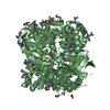
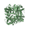
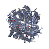
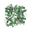
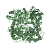
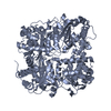
| Title | Crystal Structure of Human Presequence Protease in Complex with Amyloid-beta (1-40) | ||||||
 Components Components |
| ||||||
 Keywords Keywords | HYDROLASE/PROTEIN BINDING / M16 metalloprotease / Alzheimer's disease / zinc metalloendoprotease / monomethyllysine / dimethyllysine / S-(dimethylarsenic)cysteine / mitochondrial matrix / HYDROLASE-PROTEIN BINDING complex | ||||||
| Function / homology |  Function and homology information Function and homology informationMitochondrial protein import / Hydrolases; Acting on peptide bonds (peptidases); Metalloendopeptidases / amyloid-beta complex / growth cone lamellipodium / cellular response to norepinephrine stimulus / collateral sprouting in absence of injury / growth cone filopodium / microglia development / Formyl peptide receptors bind formyl peptides and many other ligands / regulation of Wnt signaling pathway ...Mitochondrial protein import / Hydrolases; Acting on peptide bonds (peptidases); Metalloendopeptidases / amyloid-beta complex / growth cone lamellipodium / cellular response to norepinephrine stimulus / collateral sprouting in absence of injury / growth cone filopodium / microglia development / Formyl peptide receptors bind formyl peptides and many other ligands / regulation of Wnt signaling pathway / axo-dendritic transport / axon midline choice point recognition / regulation of synapse structure or activity / hippocampal neuron apoptotic process / astrocyte activation involved in immune response / NMDA selective glutamate receptor signaling pathway / regulation of spontaneous synaptic transmission / mating behavior / : / growth factor receptor binding / peptidase activator activity / Insertion of tail-anchored proteins into the endoplasmic reticulum membrane / positive regulation of amyloid fibril formation / Golgi-associated vesicle / PTB domain binding / astrocyte projection / Lysosome Vesicle Biogenesis / Deregulated CDK5 triggers multiple neurodegenerative pathways in Alzheimer's disease models / neuron remodeling / nuclear envelope lumen / TRAF6 mediated NF-kB activation / dendrite development / positive regulation of protein metabolic process / signaling receptor activator activity / negative regulation of long-term synaptic potentiation / Advanced glycosylation endproduct receptor signaling / transition metal ion binding / The NLRP3 inflammasome / modulation of excitatory postsynaptic potential / regulation of multicellular organism growth / main axon / intracellular copper ion homeostasis / ECM proteoglycans / response to insulin-like growth factor stimulus / positive regulation of T cell migration / regulation of presynapse assembly / neuronal dense core vesicle / Purinergic signaling in leishmaniasis infection / cellular response to manganese ion / positive regulation of chemokine production / Notch signaling pathway / swimming behavior / neuron projection maintenance / extracellular matrix organization / clathrin-coated pit / positive regulation of mitotic cell cycle / axonogenesis / Mitochondrial protein degradation / positive regulation of calcium-mediated signaling / ionotropic glutamate receptor signaling pathway / platelet alpha granule lumen / astrocyte activation / response to interleukin-1 / cellular response to cAMP / regulation of neuron apoptotic process / cellular response to copper ion / positive regulation of glycolytic process / endosome lumen / trans-Golgi network membrane / positive regulation of interleukin-1 beta production / protein serine/threonine kinase binding / dendritic shaft / positive regulation of long-term synaptic potentiation / learning / central nervous system development / Post-translational protein phosphorylation / adult locomotory behavior / serine-type endopeptidase inhibitor activity / locomotory behavior / microglial cell activation / cellular response to nerve growth factor stimulus / protein processing / metalloendopeptidase activity / positive regulation of non-canonical NF-kappaB signal transduction / TAK1-dependent IKK and NF-kappa-B activation / synapse organization / visual learning / recycling endosome / positive regulation of interleukin-6 production / positive regulation of JNK cascade / Golgi lumen / regulation of long-term neuronal synaptic plasticity / response to lead ion / cognition / Regulation of Insulin-like Growth Factor (IGF) transport and uptake by Insulin-like Growth Factor Binding Proteins (IGFBPs) / cellular response to amyloid-beta / enzyme activator activity / endocytosis / metallopeptidase activity / neuron projection development Similarity search - Function | ||||||
| Biological species |  Homo sapiens (human) Homo sapiens (human) | ||||||
| Method |  X-RAY DIFFRACTION / SYNCHROTRON / molecular replacement/SAD / Resolution: 2.704 Å X-RAY DIFFRACTION / SYNCHROTRON / molecular replacement/SAD / Resolution: 2.704 Å | ||||||
 Authors Authors | King, J.V. / Liang, W.G. / Tang, W.J. | ||||||
 Citation Citation | Journal: Structure / Year: 2014 Title: Molecular basis of substrate recognition and degradation by human presequence protease. Authors: King, J.V. / Liang, W.G. / Scherpelz, K.P. / Schilling, A.B. / Meredith, S.C. / Tang, W.J. | ||||||
| History |
|
- Structure visualization
| Structure viewer | Molecule: MolmilJmol/JSmol |
|---|
- Downloads & links
-Download
| PDBx/mmCIF format | 4nge.cif.gz | 418.9 KB | Display | PDBx/mmCIF format |
|---|---|---|---|---|
| PDB format | pdb4nge.ent.gz | 331.3 KB | Display | PDB format |
| PDBx/mmJSON format | 4nge.json.gz | Tree view | PDBx/mmJSON format | |
| Others |  Other downloads Other downloads |
-Validation report
| Arichive directory | https://data.pdbj.org/pub/pdb/validation_reports/ng/4ngeftp://data.pdbj.org/pub/pdb/validation_reports/ng/4nge | HTTPS FTP |
|---|
-Related structure data


-Links
 PDBj
PDBj


- Assembly
| Deposited unit | 
| ||||||||
|---|---|---|---|---|---|---|---|---|---|
| 1 | 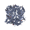
| ||||||||
| 2 | 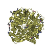
| ||||||||
| Unit cell |
|
-Components
-Presequence protease, ... , 2 types, 2 molecules AD
| #1: Protein | Mass: 115762.133 Da / Num. of mol.: 1 / Fragment: UNP residues 33-1037 / Mutation: E107Q Source method: isolated from a genetically manipulated source Source: (gene. exp.) Homo sapiens (human) / Gene: KIAA1104, MP1, PITRM1 / Plasmid: pProExH6 / Production host:  References: UniProt: Q5JRX3, Hydrolases; Acting on peptide bonds (peptidases); Metalloendopeptidases |
|---|---|
| #4: Protein | Mass: 115572.414 Da / Num. of mol.: 1 / Fragment: SEE REMARK 999 Source method: isolated from a genetically manipulated source Source: (synth.) Homo sapiens (human) / References: UniProt: Q5JRX3 |
-Beta-amyloid protein ... , 2 types, 4 molecules BECF
| #2: Protein/peptide | Mass: 4335.852 Da / Num. of mol.: 2 / Fragment: UNP residues 572-711 / Source method: obtained synthetically / Source: (synth.) Homo sapiens (human) / References: UniProt: P05067#3: Protein/peptide | Mass: 613.749 Da / Num. of mol.: 2 / Fragment: UNP residues 33-1037 / Mutation: E107Q / Source method: obtained synthetically / Source: (gene. exp.) Homo sapiens (human) / Gene: KIAA1104, MP1, PITRM1 / Plasmid: pProExH6 / Production host: References: Hydrolases; Acting on peptide bonds (peptidases); Metalloendopeptidases |
|---|
-Non-polymers , 4 types, 47 molecules 


| #5: Chemical |  Mass: 65.409 Da / Num. of mol.: 2 / Source method: obtained synthetically / Formula: Zn Mass: 65.409 Da / Num. of mol.: 2 / Source method: obtained synthetically / Formula: Zn#6: Chemical |  Mass: 92.094 Da / Num. of mol.: 3 / Source method: obtained synthetically / Formula: C3H8O3 Mass: 92.094 Da / Num. of mol.: 3 / Source method: obtained synthetically / Formula: C3H8O3#7: Chemical |  Mass: 59.044 Da / Num. of mol.: 2 / Source method: obtained synthetically / Formula: C2H3O2 Mass: 59.044 Da / Num. of mol.: 2 / Source method: obtained synthetically / Formula: C2H3O2#8: Water | ChemComp-HOH / | Mass: 18.015 Da / Num. of mol.: 40 / Source method: isolated from a natural source / Formula: H2O |
|---|
-Details
| Has protein modification | Y |
|---|---|
| Sequence details | I328V, A397V, AND Q1037R IN CHAINS A AND D ARE NATURAL VARIANTS. CHAINS C AND F ARE IDENTICAL TO ...I328V, A397V, AND Q1037R IN CHAINS A AND D ARE NATURAL VARIANTS. CHAINS C AND F ARE IDENTICAL TO CHAINS B AND E, BUT THE IDENTITIES |
-Experimental details
-Experiment
| Experiment | Method: X-RAY DIFFRACTION / Number of used crystals: 1 |
|---|
- Sample preparation
| Crystal | Density Matthews: 2.78 Å3/Da / Density % sol: 55.71 % |
|---|---|
| Crystal grow | Temperature: 291.15 K / Method: vapor diffusion, hanging drop / pH: 6.5 Details: 15.2% w/v PEG8000, 15 mM TCEP, 80 mM sodium cacodylate, pH 6.5, 160 mM calcium acetate, 20% v/v glycerol, VAPOR DIFFUSION, HANGING DROP, temperature 291.15K |
-Data collection
| Diffraction | Mean temperature: 113.5 K |
|---|---|
| Diffraction source | Source: SYNCHROTRON / Site: APS  / Beamline: 19-ID / Wavelength: 1.045 Å / Beamline: 19-ID / Wavelength: 1.045 Å |
| Detector | Type: ADSC QUANTUM 315r / Detector: CCD / Date: Jun 11, 2013 |
| Radiation | Monochromator: Rosenbaum-Rock high-resolution double-crystal Si(111) Protocol: SINGLE WAVELENGTH / Monochromatic (M) / Laue (L): M / Scattering type: x-ray |
| Radiation wavelength | Wavelength: 1.045 Å / Relative weight: 1 |
| Reflection | Resolution: 2.7→50 Å / Num. all: 72669 / Num. obs: 67540 / % possible obs: 99.1 % / Observed criterion σ(F): 0 / Observed criterion σ(I): -3 / Redundancy: 2.9 % / Biso Wilson estimate: 36.28 Å2 / Rsym value: 0.18 / Net I/σ(I): 8.69 |
| Reflection shell | Resolution: 2.7→2.75 Å / Redundancy: 2.4 % / Mean I/σ(I) obs: 1.75 / Num. unique all: 3557 / Rsym value: 0.597 / % possible all: 98.2 |
- Processing
| Software |
| ||||||||||||||||||||||||||||||||||||||||||||||||||||||||||||||||||||||||||||||||||||||||||||||||||||||||||||||||||||||||||||||||||||||||||||||||||||||||||||||||||||||||||||||||||||||||||||||||||||
|---|---|---|---|---|---|---|---|---|---|---|---|---|---|---|---|---|---|---|---|---|---|---|---|---|---|---|---|---|---|---|---|---|---|---|---|---|---|---|---|---|---|---|---|---|---|---|---|---|---|---|---|---|---|---|---|---|---|---|---|---|---|---|---|---|---|---|---|---|---|---|---|---|---|---|---|---|---|---|---|---|---|---|---|---|---|---|---|---|---|---|---|---|---|---|---|---|---|---|---|---|---|---|---|---|---|---|---|---|---|---|---|---|---|---|---|---|---|---|---|---|---|---|---|---|---|---|---|---|---|---|---|---|---|---|---|---|---|---|---|---|---|---|---|---|---|---|---|---|---|---|---|---|---|---|---|---|---|---|---|---|---|---|---|---|---|---|---|---|---|---|---|---|---|---|---|---|---|---|---|---|---|---|---|---|---|---|---|---|---|---|---|---|---|---|---|---|---|
| Refinement | Method to determine structure: molecular replacement/SAD / Resolution: 2.704→39.621 Å / SU ML: 0.26 / σ(F): 1.34 / Phase error: 24.7 / Stereochemistry target values: MLHL Details: THE PRESENCE IN THE ASYMMETRIC UNIT OF TWO PROTEINS (CHAINS A AND D) WITH DISTINCT PATTERNS OF SIDE CHAIN MODIFICATION REPRESENTS THE BEST FIT TO THE ELECTRON DENSITY AND IS NOT DERIVED FROM ...Details: THE PRESENCE IN THE ASYMMETRIC UNIT OF TWO PROTEINS (CHAINS A AND D) WITH DISTINCT PATTERNS OF SIDE CHAIN MODIFICATION REPRESENTS THE BEST FIT TO THE ELECTRON DENSITY AND IS NOT DERIVED FROM THE CO-CRYSTALLIZATION OF TWO CHEMICALLY DISTINCT PROTEINS.
| ||||||||||||||||||||||||||||||||||||||||||||||||||||||||||||||||||||||||||||||||||||||||||||||||||||||||||||||||||||||||||||||||||||||||||||||||||||||||||||||||||||||||||||||||||||||||||||||||||||
| Solvent computation | Shrinkage radii: 0.9 Å / VDW probe radii: 1.11 Å / Solvent model: FLAT BULK SOLVENT MODEL | ||||||||||||||||||||||||||||||||||||||||||||||||||||||||||||||||||||||||||||||||||||||||||||||||||||||||||||||||||||||||||||||||||||||||||||||||||||||||||||||||||||||||||||||||||||||||||||||||||||
| Displacement parameters | Biso mean: 44.63 Å2 | ||||||||||||||||||||||||||||||||||||||||||||||||||||||||||||||||||||||||||||||||||||||||||||||||||||||||||||||||||||||||||||||||||||||||||||||||||||||||||||||||||||||||||||||||||||||||||||||||||||
| Refinement step | Cycle: LAST / Resolution: 2.704→39.621 Å
| ||||||||||||||||||||||||||||||||||||||||||||||||||||||||||||||||||||||||||||||||||||||||||||||||||||||||||||||||||||||||||||||||||||||||||||||||||||||||||||||||||||||||||||||||||||||||||||||||||||
| Refine LS restraints |
| ||||||||||||||||||||||||||||||||||||||||||||||||||||||||||||||||||||||||||||||||||||||||||||||||||||||||||||||||||||||||||||||||||||||||||||||||||||||||||||||||||||||||||||||||||||||||||||||||||||
| LS refinement shell |
|
