 Movie
Movie Controller

[English] 日本語
 Yorodumi
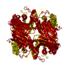
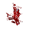
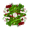
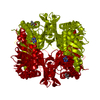
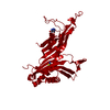
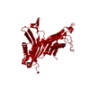
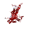
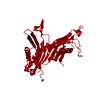
- PDB-4n9v: High resolution x-ray structure of urate oxidase in complex with ... -
+ Open data

- Basic information
| Entry | Database: PDB / ID: 4n9v | ||||||
|---|---|---|---|---|---|---|---|
| Title | High resolution x-ray structure of urate oxidase in complex with 8-azaxanthine | ||||||
 Components Components | Uricase | ||||||
 Keywords Keywords | OXIDOREDUCTASE / urate oxidase / uricase | ||||||
| Function / homology |  Function and homology information Function and homology informationfactor-independent urate hydroxylase / urate oxidase activity / purine nucleobase catabolic process / urate catabolic process / peroxisome Similarity search - Function | ||||||
| Biological species |  | ||||||
| Method |  X-RAY DIFFRACTION / SYNCHROTRON / MOLECULAR REPLACEMENT / Resolution: 1.1 Å X-RAY DIFFRACTION / SYNCHROTRON / MOLECULAR REPLACEMENT / Resolution: 1.1 Å | ||||||
 Authors Authors | Oksanen, E. / Blakeley, M.P. / Budayova-Spano, M. | ||||||
 Citation Citation | Journal: Plos One / Year: 2014 Title: The neutron structure of urate oxidase resolves a long-standing mechanistic conundrum and reveals unexpected changes in protonation. Authors: Oksanen, E. / Blakeley, M.P. / El-Hajji, M. / Ryde, U. / Budayova-Spano, M. #1: Journal: J R Soc Interface / Year: 2009 Title: Large crystal growth by thermal control allows combined X-ray and neutron crystallographic studies to elucidate the protonation states in Aspergillus flavus urate oxidase. Authors: Oksanen, E. / Blakeley, M.P. / Bonnete, F. / Dauvergne, M.T. / Dauvergne, F. / Budayova-Spano, M. | ||||||
| History |
|
- Structure visualization
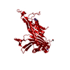
| Structure viewer | Molecule: MolmilJmol/JSmol |
|---|
- Downloads & links
-Download
| PDBx/mmCIF format | 4n9v.cif.gz | 238.3 KB | Display | PDBx/mmCIF format |
|---|---|---|---|---|
| PDB format | pdb4n9v.ent.gz | 187.4 KB | Display | PDB format |
| PDBx/mmJSON format | 4n9v.json.gz | Tree view | PDBx/mmJSON format | |
| Others |  Other downloads Other downloads |
-Validation report
| Arichive directory | https://data.pdbj.org/pub/pdb/validation_reports/n9/4n9vftp://data.pdbj.org/pub/pdb/validation_reports/n9/4n9v | HTTPS FTP |
|---|
-Related structure data
| Related structure data |  4n3mC  4n9mC  4n9sC  2ibaS S: Starting model for refinement C: citing same article ( |
|---|---|
| Similar structure data |

-Links
 PDBj
PDBj


- Assembly
| Deposited unit | 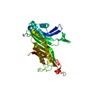
| ||||||||
|---|---|---|---|---|---|---|---|---|---|
| 1 | 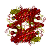
| ||||||||
| Unit cell |
| ||||||||
| Components on special symmetry positions |
|
-Components
-Protein , 1 types, 1 molecules A
| #1: Protein | Mass: 34183.590 Da / Num. of mol.: 1 Source method: isolated from a genetically manipulated source Source: (gene. exp.)  References: UniProt: Q00511, factor-independent urate hydroxylase |
|---|
-Non-polymers , 6 types, 540 molecules 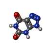


| #2: Chemical |  Mass: 153.099 Da / Num. of mol.: 3 / Source method: obtained synthetically / Formula: C4H3N5O2 Mass: 153.099 Da / Num. of mol.: 3 / Source method: obtained synthetically / Formula: C4H3N5O2#3: Chemical | ChemComp-NA / |  Mass: 22.990 Da / Num. of mol.: 1 / Source method: obtained synthetically / Formula: Na Mass: 22.990 Da / Num. of mol.: 1 / Source method: obtained synthetically / Formula: Na#4: Chemical |  Mass: 65.409 Da / Num. of mol.: 2 / Source method: obtained synthetically / Formula: Zn Mass: 65.409 Da / Num. of mol.: 2 / Source method: obtained synthetically / Formula: Zn#5: Chemical | ChemComp-GOL / |  Mass: 92.094 Da / Num. of mol.: 1 / Source method: obtained synthetically / Formula: C3H8O3 Mass: 92.094 Da / Num. of mol.: 1 / Source method: obtained synthetically / Formula: C3H8O3#6: Chemical | ChemComp-CL / |  Mass: 35.453 Da / Num. of mol.: 1 / Source method: obtained synthetically / Formula: Cl Mass: 35.453 Da / Num. of mol.: 1 / Source method: obtained synthetically / Formula: Cl#7: Chemical | ChemComp-DOD / |  Mass: 18.015 Da / Num. of mol.: 532 / Source method: isolated from a natural source / Formula: D2O Mass: 18.015 Da / Num. of mol.: 532 / Source method: isolated from a natural source / Formula: D2O |
|---|
-Details
| Has protein modification | Y |
|---|
-Experimental details
-Experiment
| Experiment | Method: X-RAY DIFFRACTION / Number of used crystals: 1 |
|---|
- Sample preparation
| Crystal | Density Matthews: 2.9 Å3/Da / Density % sol: 57.57 % |
|---|---|
| Crystal grow | Temperature: 291 K / pH: 8.5 Details: 5 % PEG 8000, 0.1 M NACL, 0.1M TRISHCL PD 8.5, 8 MG/ML URATE OXIDASE, temperature-controlled batch, temperature 291K |
-Data collection
| Diffraction | Mean temperature: 100 K |
|---|---|
| Diffraction source | Source: SYNCHROTRON / Site: ESRF  / Beamline: ID14-2 / Wavelength: 0.933 Å / Beamline: ID14-2 / Wavelength: 0.933 Å |
| Detector | Type: ADSC QUANTUM 4r / Detector: CCD / Date: Dec 4, 2007 |
| Radiation | Monochromator: diamond / Protocol: SINGLE WAVELENGTH / Monochromatic (M) / Laue (L): M / Scattering type: x-ray |
| Radiation wavelength | Wavelength: 0.933 Å / Relative weight: 1 |
| Reflection | Resolution: 1.1→50 Å / Num. all: 158812 / Num. obs: 158812 / % possible obs: 99.1 % / Observed criterion σ(F): 0 / Observed criterion σ(I): 0 / Rmerge(I) obs: 0.049 / Net I/σ(I): 11.94 |
| Reflection shell | Resolution: 1.1→1.13 Å / Rmerge(I) obs: 0.413 / Mean I/σ(I) obs: 2.93 / % possible all: 99.7 |
- Processing
| Software |
| |||||||||||||||||||||||||||||||||||||||||||||||||||||||||||||||||||||||||||||
|---|---|---|---|---|---|---|---|---|---|---|---|---|---|---|---|---|---|---|---|---|---|---|---|---|---|---|---|---|---|---|---|---|---|---|---|---|---|---|---|---|---|---|---|---|---|---|---|---|---|---|---|---|---|---|---|---|---|---|---|---|---|---|---|---|---|---|---|---|---|---|---|---|---|---|---|---|---|---|
| Refinement | Method to determine structure: MOLECULAR REPLACEMENT Starting model: PDB ENTRY 2IBA Resolution: 1.1→35.162 Å / SU ML: 0.12 / σ(F): 0 / Phase error: 13.83 / Stereochemistry target values: ML Details: EXPLICIT HYDROGENS DERIVED FROM THE NEUTRON STRUCTURE WERE USED IN RIDING POSITIONS IN THIS STRUCTURE, AND PROBABLY THESE RESIDUES HAVE DIFFERENT LEUCINE ROTAMERS (A244 AND A268) THAN IN THE NEUTRON STRUCTURE.
| |||||||||||||||||||||||||||||||||||||||||||||||||||||||||||||||||||||||||||||
| Solvent computation | Shrinkage radii: 0.9 Å / VDW probe radii: 1.11 Å / Solvent model: FLAT BULK SOLVENT MODEL / Bsol: 46.618 Å2 / ksol: 0.404 e/Å3 | |||||||||||||||||||||||||||||||||||||||||||||||||||||||||||||||||||||||||||||
| Displacement parameters |
| |||||||||||||||||||||||||||||||||||||||||||||||||||||||||||||||||||||||||||||
| Refinement step | Cycle: LAST / Resolution: 1.1→35.162 Å
| |||||||||||||||||||||||||||||||||||||||||||||||||||||||||||||||||||||||||||||
| Refine LS restraints |
| |||||||||||||||||||||||||||||||||||||||||||||||||||||||||||||||||||||||||||||
| LS refinement shell |
|
