 Movie
Movie Controller

[English] 日本語
 Yorodumi
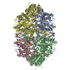
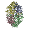
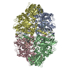
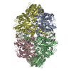
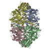
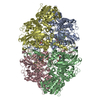
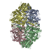
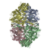
- PDB-6ztx: Crystal Structure of catalase HPII from Escherichia coli (serendi... -
+ Open data

- Basic information
| Entry | Database: PDB / ID: 6ztx | ||||||||||||
|---|---|---|---|---|---|---|---|---|---|---|---|---|---|
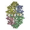
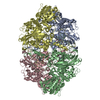
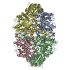
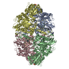
| Title | Crystal Structure of catalase HPII from Escherichia coli (serendipitously crystallized) | ||||||||||||
 Components Components | Catalase HPII | ||||||||||||
 Keywords Keywords | OXIDOREDUCTASE / catalase / hydrogen-peroxide / heme / iron / oxidative stress / artifact crystallization / impurities / contaminations | ||||||||||||
| Function / homology |  Function and homology information Function and homology informationcatalase / hyperosmotic response / catalase activity / hydrogen peroxide catabolic process / response to oxidative stress / iron ion binding / heme binding / DNA damage response / identical protein binding / cytosol / cytoplasm Similarity search - Function | ||||||||||||
| Biological species |  | ||||||||||||
| Method |  X-RAY DIFFRACTION / SYNCHROTRON / MOLECULAR REPLACEMENT / Resolution: 1.3 Å X-RAY DIFFRACTION / SYNCHROTRON / MOLECULAR REPLACEMENT / Resolution: 1.3 Å | ||||||||||||
 Authors Authors | Grzechowiak, M. / Sekula, B. / Ruszkowski, M. | ||||||||||||
| Funding support |  Poland, Poland,  United States, 3items United States, 3items
| ||||||||||||
 Citation Citation | Journal: Acta Biochim.Pol. / Year: 2021 Title: Serendipitous crystallization of E. coli HPII catalase, a sequel to "the tale usually not told". Authors: Grzechowiak, M. / Sekula, B. / Jaskolski, M. / Ruszkowski, M. | ||||||||||||
| History |
|
- Structure visualization
| Structure viewer | Molecule: MolmilJmol/JSmol |
|---|
- Downloads & links
-Download
| PDBx/mmCIF format | 6ztx.cif.gz | 1.3 MB | Display | PDBx/mmCIF format |
|---|---|---|---|---|
| PDB format | pdb6ztx.ent.gz | 1 MB | Display | PDB format |
| PDBx/mmJSON format | 6ztx.json.gz | Tree view | PDBx/mmJSON format | |
| Others |  Other downloads Other downloads |
-Validation report
| Arichive directory | https://data.pdbj.org/pub/pdb/validation_reports/zt/6ztxftp://data.pdbj.org/pub/pdb/validation_reports/zt/6ztx | HTTPS FTP |
|---|
-Related structure data
| Related structure data |  6ztvC  6ztwC  1ggeS S: Starting model for refinement C: citing same article ( |
|---|---|
| Similar structure data |


-Links
 PDBj
PDBj

- Assembly
| Deposited unit | 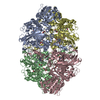
| ||||||||||||
|---|---|---|---|---|---|---|---|---|---|---|---|---|---|
| 1 |
| ||||||||||||
| Unit cell |
|
-Components
| #1: Protein | Mass: 84144.148 Da / Num. of mol.: 4 / Mutation: R37S, S99D, K372N, R521S / Source method: isolated from a natural source / Source: (natural) #2: Chemical | ChemComp-GOL /   Mass: 92.094 Da / Num. of mol.: 4 / Source method: isolated from a natural source / Formula: C3H8O3 Mass: 92.094 Da / Num. of mol.: 4 / Source method: isolated from a natural source / Formula: C3H8O3#3: Chemical | ChemComp-EDO /   Mass: 62.068 Da / Num. of mol.: 7 / Source method: obtained synthetically / Formula: C2H6O2 Mass: 62.068 Da / Num. of mol.: 7 / Source method: obtained synthetically / Formula: C2H6O2#4: Chemical | ChemComp-HDD /   Mass: 632.487 Da / Num. of mol.: 4 / Source method: obtained synthetically / Formula: C34H32FeN4O5 / Feature type: SUBJECT OF INVESTIGATION Mass: 632.487 Da / Num. of mol.: 4 / Source method: obtained synthetically / Formula: C34H32FeN4O5 / Feature type: SUBJECT OF INVESTIGATION#5: Water | ChemComp-HOH / |  Mass: 18.015 Da / Num. of mol.: 3309 / Source method: isolated from a natural source / Formula: H2O Mass: 18.015 Da / Num. of mol.: 3309 / Source method: isolated from a natural source / Formula: H2OHas ligand of interest | Y | |
|---|
-Experimental details
-Experiment
| Experiment | Method: X-RAY DIFFRACTION / Number of used crystals: 1 |
|---|
- Sample preparation
| Crystal | Density Matthews: 2.18 Å3/Da / Density % sol: 43.56 % |
|---|---|
| Crystal grow | Temperature: 292 K / Method: vapor diffusion, sitting drop / pH: 8.5 Details: 0.2 M lithium sulfate monohydrate, 0.1 M Tris pH 8.5, 25% w/v polyethylene glycol 3350 |
-Data collection
| Diffraction | Mean temperature: 100 K / Serial crystal experiment: N |
|---|---|
| Diffraction source | Source: SYNCHROTRON / Site: APS / Beamline: 22-ID / Wavelength: 1 Å |
| Detector | Type: DECTRIS EIGER X 16M / Detector: PIXEL / Date: Oct 11, 2018 |
| Radiation | Protocol: SINGLE WAVELENGTH / Monochromatic (M) / Laue (L): M / Scattering type: x-ray |
| Radiation wavelength | Wavelength: 1 Å / Relative weight: 1 |
| Reflection | Resolution: 1.3→50 Å / Num. obs: 670790 / % possible obs: 98.3 % / Redundancy: 4.84 % / Biso Wilson estimate: 11.32 Å2 / CC1/2: 0.998 / Rmerge(I) obs: 0.102 / Rrim(I) all: 0.115 / Net I/σ(I): 11.78 |
| Reflection shell | Resolution: 1.3→1.38 Å / Redundancy: 4.86 % / Rmerge(I) obs: 1.101 / Mean I/σ(I) obs: 2 / Num. unique obs: 106698 / CC1/2: 0.59 / Rrim(I) all: 1.236 / % possible all: 96.9 |
- Processing
| Software |
| ||||||||||||||||||||||||
|---|---|---|---|---|---|---|---|---|---|---|---|---|---|---|---|---|---|---|---|---|---|---|---|---|---|
| Refinement | Method to determine structure: MOLECULAR REPLACEMENT Starting model: 1gge Resolution: 1.3→44.76 Å / SU ML: 0.1299 / Cross valid method: FREE R-VALUE / σ(F): 0.69 / Phase error: 15.797 Stereochemistry target values: GeoStd + Monomer Library + CDL v1.2
| ||||||||||||||||||||||||
| Solvent computation | Shrinkage radii: 0.9 Å / VDW probe radii: 1.11 Å / Solvent model: FLAT BULK SOLVENT MODEL | ||||||||||||||||||||||||
| Displacement parameters | Biso mean: 16.76 Å2 | ||||||||||||||||||||||||
| Refinement step | Cycle: LAST / Resolution: 1.3→44.76 Å
| ||||||||||||||||||||||||
| Refine LS restraints |
|
