 Movie
Movie Controller


+ Open data

- Basic information
| Entry | Database: PDB / ID: 1qws | ||||||
|---|---|---|---|---|---|---|---|
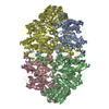
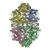
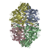
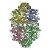
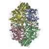
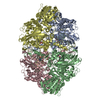
| Title | Structure of the D181N variant of catalase HPII from E. coli | ||||||
 Components Components | Catalase HPII | ||||||
 Keywords Keywords | OXIDOREDUCTASE / beta barrel / heme b | ||||||
| Function / homology |  Function and homology information Function and homology informationcatalase / catalase activity / hyperosmotic response / hydrogen peroxide catabolic process / response to oxidative stress / iron ion binding / heme binding / DNA damage response / identical protein binding / cytoplasm / cytosol Similarity search - Function | ||||||
| Biological species |  | ||||||
| Method |  X-RAY DIFFRACTION / SYNCHROTRON / MOLECULAR REPLACEMENT / Resolution: 1.9 Å X-RAY DIFFRACTION / SYNCHROTRON / MOLECULAR REPLACEMENT / Resolution: 1.9 Å | ||||||
 Authors Authors | Chelikani, P. / Carpena, X. / Fita, I. / Loewen, P.C. | ||||||
 Citation Citation | Journal: J.Biol.Chem. / Year: 2003 Title: An electrical potential in the access channel of catalases enhances catalysis Authors: Chelikani, P. / Carpena, X. / Fita, I. / Loewen, P.C. | ||||||
| History |
|
- Structure visualization
| Structure viewer | Molecule: MolmilJmol/JSmol |
|---|
- Downloads & links
-Download
| PDBx/mmCIF format | 1qws.cif.gz | 637.6 KB | Display | PDBx/mmCIF format |
|---|---|---|---|---|
| PDB format | pdb1qws.ent.gz | 518.1 KB | Display | PDB format |
| PDBx/mmJSON format | 1qws.json.gz | Tree view | PDBx/mmJSON format | |
| Others |  Other downloads Other downloads |
-Validation report
| Arichive directory | https://data.pdbj.org/pub/pdb/validation_reports/qw/1qwsftp://data.pdbj.org/pub/pdb/validation_reports/qw/1qws | HTTPS FTP |
|---|
-Related structure data
| Related structure data | 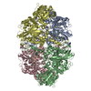 1p7yC  1p7zC 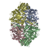 1p80C  1p81C  1ggeS C: citing same article ( S: Starting model for refinement |
|---|---|
| Similar structure data |


-Links
 PDBj
PDBj

- Assembly
| Deposited unit | 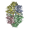
| ||||||||||||||||||||||||||||||
|---|---|---|---|---|---|---|---|---|---|---|---|---|---|---|---|---|---|---|---|---|---|---|---|---|---|---|---|---|---|---|---|
| 1 |
| ||||||||||||||||||||||||||||||
| Unit cell |
| ||||||||||||||||||||||||||||||
| Noncrystallographic symmetry (NCS) | NCS domain:
NCS domain segments: Component-ID: 1 / Ens-ID: 1 / Beg auth comp-ID: ASP / Beg label comp-ID: ASP / End auth comp-ID: ALA / End label comp-ID: ALA / Refine code: 6 / Auth seq-ID: 27 - 753 / Label seq-ID: 27 - 753
|
-Components
| #1: Protein | Mass: 84270.469 Da / Num. of mol.: 4 / Mutation: D181N Source method: isolated from a genetically manipulated source Source: (gene. exp.) #2: Chemical | ChemComp-HEM /   Mass: 616.487 Da / Num. of mol.: 4 / Source method: obtained synthetically / Formula: C34H32FeN4O4 Mass: 616.487 Da / Num. of mol.: 4 / Source method: obtained synthetically / Formula: C34H32FeN4O4#3: Water | ChemComp-HOH / |  Mass: 18.015 Da / Num. of mol.: 3033 / Source method: isolated from a natural source / Formula: H2O Mass: 18.015 Da / Num. of mol.: 3033 / Source method: isolated from a natural source / Formula: H2O |
|---|
-Experimental details
-Experiment
| Experiment | Method: X-RAY DIFFRACTION / Number of used crystals: 1 |
|---|
- Sample preparation
| Crystal | Density Matthews: 2.12 Å3/Da / Density % sol: 41.87 % |
|---|---|
| Crystal grow | Temperature: 293 K / Method: vapor diffusion, hanging drop / pH: 9 Details: 15% PEG 3350, 1.6M LiCl, 0.1M Tris, pH 9.0, VAPOR DIFFUSION, HANGING DROP, temperature 293K |
-Data collection
| Diffraction | Mean temperature: 100 K |
|---|---|
| Diffraction source | Source: SYNCHROTRON / Site: ESRF  / Beamline: BM14 / Wavelength: 0.9763 Å / Beamline: BM14 / Wavelength: 0.9763 Å |
| Detector | Type: MARRESEARCH / Detector: CCD / Date: Jul 8, 2003 |
| Radiation | Monochromator: Si 111 Channel / Protocol: SINGLE WAVELENGTH / Monochromatic (M) / Laue (L): M / Scattering type: x-ray |
| Radiation wavelength | Wavelength: 0.9763 Å / Relative weight: 1 |
| Reflection | Resolution: 1.9→27.81 Å / Num. all: 218818 / Num. obs: 218251 / % possible obs: 99.5 % / Observed criterion σ(F): 0 / Observed criterion σ(I): 0 / Rsym value: 0.097 / Net I/σ(I): 7.2 |
| Reflection shell | Resolution: 1.9→1.99 Å / Num. unique all: 15850 / % possible all: 97.3 |
- Processing
| Software |
| ||||||||||||||||||||||||||||||||||||||||||||||||||||||||||||||||||||||
|---|---|---|---|---|---|---|---|---|---|---|---|---|---|---|---|---|---|---|---|---|---|---|---|---|---|---|---|---|---|---|---|---|---|---|---|---|---|---|---|---|---|---|---|---|---|---|---|---|---|---|---|---|---|---|---|---|---|---|---|---|---|---|---|---|---|---|---|---|---|---|---|
| Refinement | Method to determine structure: MOLECULAR REPLACEMENT Starting model: PDB ENTRY 1GGE Resolution: 1.9→28 Å / Cor.coef. Fo:Fc: 0.96 / Cor.coef. Fo:Fc free: 0.931 / SU B: 3.319 / SU ML: 0.097 / Cross valid method: THROUGHOUT / σ(F): 0 / σ(I): 0 / ESU R: 0.161 / ESU R Free: 0.151 / Stereochemistry target values: MAXIMUM LIKELIHOOD
| ||||||||||||||||||||||||||||||||||||||||||||||||||||||||||||||||||||||
| Solvent computation | Ion probe radii: 0.8 Å / Shrinkage radii: 0.8 Å / VDW probe radii: 1.4 Å / Solvent model: BABINET MODEL WITH MASK | ||||||||||||||||||||||||||||||||||||||||||||||||||||||||||||||||||||||
| Displacement parameters | Biso mean: 20.472 Å2
| ||||||||||||||||||||||||||||||||||||||||||||||||||||||||||||||||||||||
| Refine analyze | Luzzati sigma a obs: 0.19 Å | ||||||||||||||||||||||||||||||||||||||||||||||||||||||||||||||||||||||
| Refinement step | Cycle: LAST / Resolution: 1.9→28 Å
| ||||||||||||||||||||||||||||||||||||||||||||||||||||||||||||||||||||||
| Refine LS restraints |
| ||||||||||||||||||||||||||||||||||||||||||||||||||||||||||||||||||||||
| Refine LS restraints NCS | Ens-ID: 1 / Number: 5745 / Refine-ID: X-RAY DIFFRACTION
| ||||||||||||||||||||||||||||||||||||||||||||||||||||||||||||||||||||||
| LS refinement shell | Resolution: 1.9→1.95 Å / Total num. of bins used: 20 /
|
