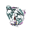
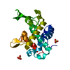
- PDB-6zqp: Structure of the Pmt2-MIR domain with bound ligands -
+
Open data
ID or keywords:
Loading...
-
Basic information
Entry
Database: PDB / ID: 6zqp
Title
Structure of the Pmt2-MIR domain with bound ligands
Components
PMT2 isoform 1
Keywords
PEPTIDE BINDING PROTEIN / carbohydrate-binding module / MIR domain / protein-O-mannosylation / beta-trefoil
Function / homology
Function and homology information
dolichyl-phosphate-mannose-protein mannosyltransferase Pmt1p-Pmt2p dimer complex / dolichyl-phosphate-mannose-protein mannosyltransferase Pmt5p-Pmt2p dimer complex / dolichyl-phosphate-mannose-protein mannosyltransferase / dolichyl-phosphate-mannose-protein mannosyltransferase activity / regulation of endoplasmic reticulum unfolded protein response / fungal-type cell wall biogenesis / protein O-linked glycosylation via mannose / protein exit from endoplasmic reticulum / protein O-linked glycosylation / membrane => GO:0016020 ...dolichyl-phosphate-mannose-protein mannosyltransferase Pmt1p-Pmt2p dimer complex / dolichyl-phosphate-mannose-protein mannosyltransferase Pmt5p-Pmt2p dimer complex / dolichyl-phosphate-mannose-protein mannosyltransferase / dolichyl-phosphate-mannose-protein mannosyltransferase activity / regulation of endoplasmic reticulum unfolded protein response / fungal-type cell wall biogenesis / protein O-linked glycosylation via mannose / protein exit from endoplasmic reticulum / protein O-linked glycosylation / membrane => GO:0016020 / ERAD pathway / endoplasmic reticulum membrane / endoplasmic reticulum Similarity search - Function
Glycosyl transferase family 39/83 / Glycosyltransferase 39-like / Protein O-mannosyl-transferase, C-terminal four TM domain / Dolichyl-phosphate-mannose-protein mannosyltransferase / C-terminal four TMM region of protein-O-mannosyltransferase / MIR motif / MIR domain / MIR domain profile. / Domain in ryanodine and inositol trisphosphate receptors and protein O-mannosyltransferases / Mir domain superfamily Similarity search - Domain/homology
In the structure databanks used in Yorodumi, some data are registered as the other names, "COVID-19 virus" and "2019-nCoV". Here are the details of the virus and the list of structure data.
Jan 31, 2019. EMDB accession codes are about to change! (news from PDBe EMDB page)
EMDB accession codes are about to change! (news from PDBe EMDB page)
The allocation of 4 digits for EMDB accession codes will soon come to an end. Whilst these codes will remain in use, new EMDB accession codes will include an additional digit and will expand incrementally as the available range of codes is exhausted. The current 4-digit format prefixed with “EMD-” (i.e. EMD-XXXX) will advance to a 5-digit format (i.e. EMD-XXXXX), and so on. It is currently estimated that the 4-digit codes will be depleted around Spring 2019, at which point the 5-digit format will come into force.
The EM Navigator/Yorodumi systems omit the EMD- prefix.
Related info.:Q: What is EMD? / ID/Accession-code notation in Yorodumi/EM Navigator
Yorodumi is a browser for structure data from EMDB, PDB, SASBDB, etc.
This page is also the successor to EM Navigator detail page, and also detail information page/front-end page for Omokage search.
The word "yorodu" (or yorozu) is an old Japanese word meaning "ten thousand". "mi" (miru) is to see.
Related info.:EMDB / PDB / SASBDB / Comparison of 3 databanks / Yorodumi Search / Aug 31, 2016. New EM Navigator & Yorodumi / Yorodumi Papers / Jmol/JSmol / Function and homology information / Changes in new EM Navigator and Yorodumi
 Movie
Movie Controller


 Open data
 Basic information
 Components
 Keywords
 Function and homology information
 X-RAY DIFFRACTION /
  Authors
 Citation
 Structure visualization
 Downloads & links
 Other downloads


 PDBj
PDBj Assembly
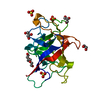


 Mass: 194.226 Da / Num. of mol.: 1 / Source method: obtained synthetically / Formula: C8H18O5 / Comment: precipitant*YM
 Mass: 92.094 Da / Num. of mol.: 3 / Source method: obtained synthetically / Formula: C3H8O3 / Feature type: SUBJECT OF INVESTIGATION
 Mass: 96.063 Da / Num. of mol.: 4 / Source method: obtained synthetically / Formula: SO4
 Mass: 18.015 Da / Num. of mol.: 280 / Source method: isolated from a natural source / Formula: H2O
 Sample preparation
 / Beamline: ID29 / Wavelength: 1.072 Å
 Processing