 Movie
Movie Controller


+ Open data

- Basic information
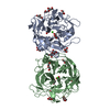
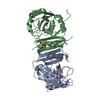
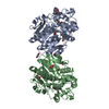
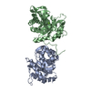
| Entry | Database: PDB / ID: 6zk4 | ||||||
|---|---|---|---|---|---|---|---|
| Title | Plant nucleoside hydrolase - ZmNRh2b with a bound adenine | ||||||
 Components Components | Pyrimidine-specific ribonucleoside hydrolase rihA | ||||||
 Keywords Keywords | HYDROLASE / Plant enzyme | ||||||
| Function / homology |  Function and homology information Function and homology informationribonucleoside catabolic process / uridine nucleosidase / inosine nucleosidase activity / uridine nucleosidase activity / purine nucleosidase activity / purine nucleoside catabolic process / protein heterodimerization activity / metal ion binding / cytosol Similarity search - Function | ||||||
| Biological species |  | ||||||
| Method |  X-RAY DIFFRACTION / SYNCHROTRON / MOLECULAR REPLACEMENT / Resolution: 1.95 Å X-RAY DIFFRACTION / SYNCHROTRON / MOLECULAR REPLACEMENT / Resolution: 1.95 Å | ||||||
 Authors Authors | Morera, S. / Vigouroux, A. / Kopecny, D. | ||||||
 Citation Citation | Journal: Plant J. / Year: 2023 Title: Plant nucleoside N-ribohydrolases: riboside binding and role in nitrogen storage mobilization. Authors: Luptakova, E. / Vigouroux, A. / Koncitikova, R. / Kopecna, M. / Zalabak, D. / Novak, O. / Salcedo Sarmiento, S. / Cavar Zeljkovic, S. / Kopecny, D.J. / von Schwartzenberg, K. / Strnad, M. / ...Authors: Luptakova, E. / Vigouroux, A. / Koncitikova, R. / Kopecna, M. / Zalabak, D. / Novak, O. / Salcedo Sarmiento, S. / Cavar Zeljkovic, S. / Kopecny, D.J. / von Schwartzenberg, K. / Strnad, M. / Spichal, L. / De Diego, N. / Kopecny, D. / Morera, S. | ||||||
| History |
|
- Structure visualization
| Structure viewer | Molecule: MolmilJmol/JSmol |
|---|
- Downloads & links
-Download
| PDBx/mmCIF format | 6zk4.cif.gz | 501.6 KB | Display | PDBx/mmCIF format |
|---|---|---|---|---|
| PDB format | pdb6zk4.ent.gz | 412.3 KB | Display | PDB format |
| PDBx/mmJSON format | 6zk4.json.gz | Tree view | PDBx/mmJSON format | |
| Others |  Other downloads Other downloads |
-Validation report
| Arichive directory | https://data.pdbj.org/pub/pdb/validation_reports/zk/6zk4ftp://data.pdbj.org/pub/pdb/validation_reports/zk/6zk4 | HTTPS FTP |
|---|
-Related structure data
| Related structure data |  6zk1C  6zk2C 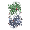 6zk3C  6zk5C  4kpoS S: Starting model for refinement C: citing same article ( |
|---|---|
| Similar structure data |


-Links
 PDBj
PDBj


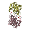
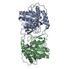
- Assembly
| Deposited unit | 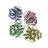
| ||||||||
|---|---|---|---|---|---|---|---|---|---|
| 1 | 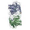
| ||||||||
| 2 | 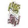
| ||||||||
| Unit cell |
|
-Components
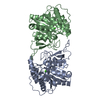
-Protein , 1 types, 4 molecules ABCD
| #1: Protein | Mass: 37122.316 Da / Num. of mol.: 4 Source method: isolated from a genetically manipulated source Source: (gene. exp.)  |
|---|
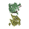
-Non-polymers , 5 types, 531 molecules 


| #2: Chemical | ChemComp-CA /  Mass: 40.078 Da / Num. of mol.: 4 / Source method: obtained synthetically / Formula: Ca Mass: 40.078 Da / Num. of mol.: 4 / Source method: obtained synthetically / Formula: Ca#3: Chemical | ChemComp-ADE /  Mass: 135.127 Da / Num. of mol.: 4 / Source method: obtained synthetically / Formula: C5H5N5 / Feature type: SUBJECT OF INVESTIGATION Mass: 135.127 Da / Num. of mol.: 4 / Source method: obtained synthetically / Formula: C5H5N5 / Feature type: SUBJECT OF INVESTIGATION#4: Chemical |  Mass: 106.120 Da / Num. of mol.: 2 / Source method: obtained synthetically / Formula: C4H10O3 Mass: 106.120 Da / Num. of mol.: 2 / Source method: obtained synthetically / Formula: C4H10O3#5: Chemical | ChemComp-EDO / |  Mass: 62.068 Da / Num. of mol.: 1 / Source method: obtained synthetically / Formula: C2H6O2 Mass: 62.068 Da / Num. of mol.: 1 / Source method: obtained synthetically / Formula: C2H6O2#6: Water | ChemComp-HOH / | Mass: 18.015 Da / Num. of mol.: 520 / Source method: isolated from a natural source / Formula: H2O |
|---|
-Details
| Has ligand of interest | Y |
|---|
-Experimental details
-Experiment
| Experiment | Method: X-RAY DIFFRACTION / Number of used crystals: 1 |
|---|
- Sample preparation
| Crystal | Density Matthews: 2.05 Å3/Da / Density % sol: 40 % |
|---|---|
| Crystal grow | Temperature: 292 K / Method: vapor diffusion, hanging drop / Details: PEG 4000, Tris |
-Data collection
| Diffraction | Mean temperature: 100 K / Serial crystal experiment: N |
|---|---|
| Diffraction source | Source: SYNCHROTRON / Site: SOLEIL  / Beamline: PROXIMA 1 / Wavelength: 0.978 Å / Beamline: PROXIMA 1 / Wavelength: 0.978 Å |
| Detector | Type: DECTRIS PILATUS3 S 6M / Detector: PIXEL / Date: Mar 19, 2016 |
| Radiation | Protocol: SINGLE WAVELENGTH / Monochromatic (M) / Laue (L): M / Scattering type: x-ray |
| Radiation wavelength | Wavelength: 0.978 Å / Relative weight: 1 |
| Reflection | Resolution: 1.95→42.86 Å / Num. obs: 86204 / % possible obs: 98.5 % / Redundancy: 3 % / CC1/2: 0.998 / Rmerge(I) obs: 0.058 / Net I/σ(I): 10.4 |
| Reflection shell | Resolution: 1.95→2.06 Å / Num. unique obs: 13693 / CC1/2: 0.628 |
- Processing
| Software |
| |||||||||||||||||||||||||||||||||||||||||||||||||||||||||||||||||||||||||||||||||||||||||||||||||||||||||||||||||||||||||||||
|---|---|---|---|---|---|---|---|---|---|---|---|---|---|---|---|---|---|---|---|---|---|---|---|---|---|---|---|---|---|---|---|---|---|---|---|---|---|---|---|---|---|---|---|---|---|---|---|---|---|---|---|---|---|---|---|---|---|---|---|---|---|---|---|---|---|---|---|---|---|---|---|---|---|---|---|---|---|---|---|---|---|---|---|---|---|---|---|---|---|---|---|---|---|---|---|---|---|---|---|---|---|---|---|---|---|---|---|---|---|---|---|---|---|---|---|---|---|---|---|---|---|---|---|---|---|---|
| Refinement | Method to determine structure: MOLECULAR REPLACEMENT Starting model: 4KPO Resolution: 1.95→42.86 Å / Cor.coef. Fo:Fc: 0.958 / Cor.coef. Fo:Fc free: 0.948 / SU R Cruickshank DPI: 0.162 / Cross valid method: THROUGHOUT / σ(F): 0 / SU R Blow DPI: 0.16 / SU Rfree Blow DPI: 0.134 / SU Rfree Cruickshank DPI: 0.137
| |||||||||||||||||||||||||||||||||||||||||||||||||||||||||||||||||||||||||||||||||||||||||||||||||||||||||||||||||||||||||||||
| Displacement parameters | Biso max: 153.38 Å2 / Biso mean: 50.74 Å2 / Biso min: 22.3 Å2
| |||||||||||||||||||||||||||||||||||||||||||||||||||||||||||||||||||||||||||||||||||||||||||||||||||||||||||||||||||||||||||||
| Refine analyze | Luzzati coordinate error obs: 0.25 Å | |||||||||||||||||||||||||||||||||||||||||||||||||||||||||||||||||||||||||||||||||||||||||||||||||||||||||||||||||||||||||||||
| Refinement step | Cycle: final / Resolution: 1.95→42.86 Å
| |||||||||||||||||||||||||||||||||||||||||||||||||||||||||||||||||||||||||||||||||||||||||||||||||||||||||||||||||||||||||||||
| Refine LS restraints |
| |||||||||||||||||||||||||||||||||||||||||||||||||||||||||||||||||||||||||||||||||||||||||||||||||||||||||||||||||||||||||||||
| LS refinement shell | Resolution: 1.95→1.96 Å / Rfactor Rfree error: 0 / Total num. of bins used: 50
| |||||||||||||||||||||||||||||||||||||||||||||||||||||||||||||||||||||||||||||||||||||||||||||||||||||||||||||||||||||||||||||
| Refinement TLS params. | Method: refined / Refine-ID: X-RAY DIFFRACTION
| |||||||||||||||||||||||||||||||||||||||||||||||||||||||||||||||||||||||||||||||||||||||||||||||||||||||||||||||||||||||||||||
| Refinement TLS group |
|
