 Movie
Movie Controller

[English] 日本語
 Yorodumi
- PDB-4c4x: Crystal structure of human bifunctional epoxide hydroxylase 2 com... -
+ Open data

- Basic information
| Entry | Database: PDB / ID: 4c4x | ||||||
|---|---|---|---|---|---|---|---|
| Title | Crystal structure of human bifunctional epoxide hydroxylase 2 complexed with C9 | ||||||
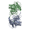
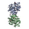
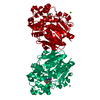
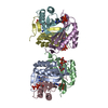
 Components Components | BIFUNCTIONAL EPOXIDE HYDROLASE 2 | ||||||
 Keywords Keywords | HYDROLASE / DRUG DESIGN / MULTIPLE BINDING MODES | ||||||
| Function / homology |  Function and homology information Function and homology informationlipid-phosphate phosphatase / 10-hydroxy-9-(phosphonooxy)octadecanoate phosphatase activity / stilbene catabolic process / Biosynthesis of maresins / epoxide metabolic process / lipid phosphatase activity / phospholipid dephosphorylation / Synthesis of epoxy (EET) and dihydroxyeicosatrienoic acids (DHET) / lysophosphatidic acid phosphatase activity / soluble epoxide hydrolase ...lipid-phosphate phosphatase / 10-hydroxy-9-(phosphonooxy)octadecanoate phosphatase activity / stilbene catabolic process / Biosynthesis of maresins / epoxide metabolic process / lipid phosphatase activity / phospholipid dephosphorylation / Synthesis of epoxy (EET) and dihydroxyeicosatrienoic acids (DHET) / lysophosphatidic acid phosphatase activity / soluble epoxide hydrolase / epoxide hydrolase activity / dephosphorylation / regulation of cholesterol metabolic process / phosphatase activity / peroxisomal matrix / toxic substance binding / cholesterol homeostasis / regulation of cell growth / Peroxisomal protein import / response to toxic substance / peroxisome / positive regulation of gene expression / magnesium ion binding / protein homodimerization activity / extracellular exosome / cytosol Similarity search - Function | ||||||
| Biological species |  HOMO SAPIENS (human) HOMO SAPIENS (human) | ||||||
| Method |  X-RAY DIFFRACTION / SYNCHROTRON / MOLECULAR REPLACEMENT / Resolution: 2.17 Å X-RAY DIFFRACTION / SYNCHROTRON / MOLECULAR REPLACEMENT / Resolution: 2.17 Å | ||||||
 Authors Authors | Pilger, J. / Mazur, A. / Monecke, P. / Schreuder, H. / Elshorst, B. / Langer, T. / Schiffer, A. / Krimm, I. / Wegstroth, M. / Lee, D. ...Pilger, J. / Mazur, A. / Monecke, P. / Schreuder, H. / Elshorst, B. / Langer, T. / Schiffer, A. / Krimm, I. / Wegstroth, M. / Lee, D. / Hessler, G. / Wendt, K.-U. / Becker, S. / Griesinger, C. | ||||||
 Citation Citation | Journal: Angew.Chem.Int.Ed.Engl. / Year: 2015 Title: A Combination of Spin Diffusion Methods for the Determination of Protein-Ligand Complex Structural Ensembles. Authors: Pilger, J. / Mazur, A. / Monecke, P. / Schreuder, H. / Elshorst, B. / Bartoschek, S. / Langer, T. / Schiffer, A. / Krimm, I. / Wegstroth, M. / Lee, D. / Hessler, G. / Wendt, K. / Becker, S. / Griesinger, C. | ||||||
| History |
|
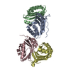
- Structure visualization
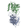
| Structure viewer | Molecule: MolmilJmol/JSmol |
|---|
- Downloads & links
-Download
| PDBx/mmCIF format | 4c4x.cif.gz | 148.7 KB | Display | PDBx/mmCIF format |
|---|---|---|---|---|
| PDB format | pdb4c4x.ent.gz | 116.3 KB | Display | PDB format |
| PDBx/mmJSON format | 4c4x.json.gz | Tree view | PDBx/mmJSON format | |
| Others |  Other downloads Other downloads |
-Validation report
| Arichive directory | https://data.pdbj.org/pub/pdb/validation_reports/c4/4c4xftp://data.pdbj.org/pub/pdb/validation_reports/c4/4c4x | HTTPS FTP |
|---|
-Related structure data
| Related structure data |  4c4yC 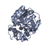 4c4zC  4yxrC  4yxsC 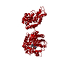 3otqS C: citing same article ( S: Starting model for refinement |
|---|---|
| Similar structure data |


-Links
 PDBj
PDBj


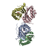
- Assembly
| Deposited unit | 
| ||||||||
|---|---|---|---|---|---|---|---|---|---|
| 1 | 
| ||||||||
| Unit cell |
|
-Components
| #1: Protein | Mass: 37370.926 Da / Num. of mol.: 2 / Fragment: EPOXIDE HYDROXYLASE DOMAIN RESIDUES 230-555 Source method: isolated from a genetically manipulated source Source: (gene. exp.) HOMO SAPIENS (human) / Production host:  References: UniProt: P34913, soluble epoxide hydrolase, lipid-phosphate phosphatase #2: Chemical |   Mass: 233.094 Da / Num. of mol.: 2 / Source method: obtained synthetically / Formula: C9H10Cl2N2O Mass: 233.094 Da / Num. of mol.: 2 / Source method: obtained synthetically / Formula: C9H10Cl2N2O#3: Water | ChemComp-HOH / |  Mass: 18.015 Da / Num. of mol.: 445 / Source method: isolated from a natural source / Formula: H2O Mass: 18.015 Da / Num. of mol.: 445 / Source method: isolated from a natural source / Formula: H2O |
|---|
-Experimental details
-Experiment
| Experiment | Method: X-RAY DIFFRACTION / Number of used crystals: 1 |
|---|
- Sample preparation
| Crystal | Density Matthews: 2.35 Å3/Da / Density % sol: 47.6 % / Description: NONE |
|---|---|
| Crystal grow | Temperature: 293 K / Method: vapor diffusion, hanging drop / pH: 8.3 Details: HANGING DROP. 1 UL PROTEIN SOLUTION (8 MG/ML SEH IN 100 MM NAPO2, PH 7.4, 5 MM DTT AND 15% GLYCEROL) WAS MIXED WITH 1 UL RESERVOIR SOLUTION (100 MM TRIS-HCL, PH 8.3, 26% PEG4000 AND 200 MM ...Details: HANGING DROP. 1 UL PROTEIN SOLUTION (8 MG/ML SEH IN 100 MM NAPO2, PH 7.4, 5 MM DTT AND 15% GLYCEROL) WAS MIXED WITH 1 UL RESERVOIR SOLUTION (100 MM TRIS-HCL, PH 8.3, 26% PEG4000 AND 200 MM LI2SO4 AND EQUILIBRATED AT 20C. CRYSTALS APPEARED IN ABOUT 2 WEEKS |
-Data collection
| Diffraction | Mean temperature: 100 K |
|---|---|
| Diffraction source | Source: SYNCHROTRON / Site: SLS  / Beamline: X06DA / Wavelength: 1 / Beamline: X06DA / Wavelength: 1 |
| Detector | Type: DECTRIS PILATUS / Detector: PIXEL / Date: Nov 30, 2012 |
| Radiation | Protocol: SINGLE WAVELENGTH / Monochromatic (M) / Laue (L): M / Scattering type: x-ray |
| Radiation wavelength | Wavelength: 1 Å / Relative weight: 1 |
| Reflection | Resolution: 2.17→44.31 Å / Num. obs: 35085 / % possible obs: 98.9 % / Observed criterion σ(I): -3 / Redundancy: 3.3 % / Biso Wilson estimate: 35.12 Å2 / Rmerge(I) obs: 0.09 / Net I/σ(I): 7.9 |
| Reflection shell | Resolution: 2.17→2.28 Å / Redundancy: 3.3 % / Rmerge(I) obs: 0.41 / Mean I/σ(I) obs: 2.4 / % possible all: 99.4 |
- Processing
| Software |
| ||||||||||||||||||||||||||||||||||||||||||||||||||||||||||||||||||||||||||||||||||||||||||||||||||||||||||||||||||
|---|---|---|---|---|---|---|---|---|---|---|---|---|---|---|---|---|---|---|---|---|---|---|---|---|---|---|---|---|---|---|---|---|---|---|---|---|---|---|---|---|---|---|---|---|---|---|---|---|---|---|---|---|---|---|---|---|---|---|---|---|---|---|---|---|---|---|---|---|---|---|---|---|---|---|---|---|---|---|---|---|---|---|---|---|---|---|---|---|---|---|---|---|---|---|---|---|---|---|---|---|---|---|---|---|---|---|---|---|---|---|---|---|---|---|---|
| Refinement | Method to determine structure: MOLECULAR REPLACEMENT Starting model: PDB ENTRY 3OTQ Resolution: 2.17→29.63 Å / Cor.coef. Fo:Fc: 0.9089 / Cor.coef. Fo:Fc free: 0.879 / SU R Cruickshank DPI: 0.304 / Cross valid method: THROUGHOUT / σ(F): 0 / SU R Blow DPI: 0.319 / SU Rfree Blow DPI: 0.211 / SU Rfree Cruickshank DPI: 0.21 Details: IDEAL-DIST CONTACT TERM CONTACT SETUP. ALL ATOMS HAVE CCP4 ATOM TYPE FROM LIBRARY. THE INHIBITOR HAS BEEN MODELED IN TWO ORIENTATIONS
| ||||||||||||||||||||||||||||||||||||||||||||||||||||||||||||||||||||||||||||||||||||||||||||||||||||||||||||||||||
| Displacement parameters | Biso mean: 38.63 Å2
| ||||||||||||||||||||||||||||||||||||||||||||||||||||||||||||||||||||||||||||||||||||||||||||||||||||||||||||||||||
| Refine analyze | Luzzati coordinate error obs: 0.287 Å | ||||||||||||||||||||||||||||||||||||||||||||||||||||||||||||||||||||||||||||||||||||||||||||||||||||||||||||||||||
| Refinement step | Cycle: LAST / Resolution: 2.17→29.63 Å
| ||||||||||||||||||||||||||||||||||||||||||||||||||||||||||||||||||||||||||||||||||||||||||||||||||||||||||||||||||
| Refine LS restraints |
| ||||||||||||||||||||||||||||||||||||||||||||||||||||||||||||||||||||||||||||||||||||||||||||||||||||||||||||||||||
| LS refinement shell | Resolution: 2.17→2.24 Å / Total num. of bins used: 17
|
