 Movie
Movie Controller


+ Open data

- Basic information
| Entry | Database: PDB / ID: 4kpn | ||||||
|---|---|---|---|---|---|---|---|
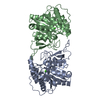
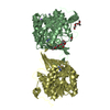
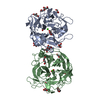
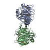
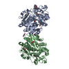
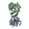
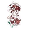
| Title | Plant nucleoside hydrolase - PpNRh1 enzyme | ||||||
 Components Components | Nucleoside N-ribohydrolase 1 | ||||||
 Keywords Keywords | HYDROLASE / IU-NRHs | ||||||
| Function / homology |  Function and homology information Function and homology informationpurine nucleosidase / inosine nucleosidase activity / purine nucleosidase activity / leaf senescence / inosine catabolic process / UDP-glucosyltransferase activity / purine nucleoside catabolic process / protein heterodimerization activity / metal ion binding / cytosol Similarity search - Function | ||||||
| Biological species |  Physcomitrella patens (plant) Physcomitrella patens (plant) | ||||||
| Method |  X-RAY DIFFRACTION / SYNCHROTRON / MOLECULAR REPLACEMENT / Resolution: 3.35 Å X-RAY DIFFRACTION / SYNCHROTRON / MOLECULAR REPLACEMENT / Resolution: 3.35 Å | ||||||
 Authors Authors | Morera, S. / Vigouroux, A. / Kopecny, D. | ||||||
 Citation Citation | Journal: Plant Physiol. / Year: 2013 Title: Structure and Function of Nucleoside Hydrolases from Physcomitrella patens and Maize Catalyzing the Hydrolysis of Purine, Pyrimidine, and Cytokinin Ribosides. Authors: Kopecna, M. / Blaschke, H. / Kopecny, D. / Vigouroux, A. / Koncitikova, R. / Novak, O. / Kotland, O. / Strnad, M. / Morera, S. / von Schwartzenberg, K. | ||||||
| History |
|
- Structure visualization
| Structure viewer | Molecule: MolmilJmol/JSmol |
|---|
- Downloads & links
-Download
| PDBx/mmCIF format | 4kpn.cif.gz | 963.2 KB | Display | PDBx/mmCIF format |
|---|---|---|---|---|
| PDB format | pdb4kpn.ent.gz | 811.3 KB | Display | PDB format |
| PDBx/mmJSON format | 4kpn.json.gz | Tree view | PDBx/mmJSON format | |
| Others |  Other downloads Other downloads |
-Validation report
| Arichive directory | https://data.pdbj.org/pub/pdb/validation_reports/kp/4kpnftp://data.pdbj.org/pub/pdb/validation_reports/kp/4kpn | HTTPS FTP |
|---|
-Related structure data
| Related structure data |  4kpoC 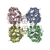 3b9xS S: Starting model for refinement C: citing same article ( |
|---|---|
| Similar structure data |


-Links
 PDBj
PDBj- Assembly
| Deposited unit | 
| ||||||||
|---|---|---|---|---|---|---|---|---|---|
| 1 | 
| ||||||||
| 2 | 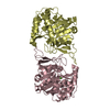
| ||||||||
| 3 | 
| ||||||||
| 4 | 
| ||||||||
| Unit cell |
|
-Components
| #1: Protein | Mass: 37474.277 Da / Num. of mol.: 8 Source method: isolated from a genetically manipulated source Source: (gene. exp.) Physcomitrella patens (plant) / Gene: NRH1 / Production host:  References: UniProt: M1FQT3, UniProt: A9TXA6*PLUS, purine nucleosidase #2: Chemical | ChemComp-CA /   Mass: 40.078 Da / Num. of mol.: 8 / Source method: obtained synthetically / Formula: Ca Mass: 40.078 Da / Num. of mol.: 8 / Source method: obtained synthetically / Formula: Ca#3: Water | ChemComp-HOH / |  Mass: 18.015 Da / Num. of mol.: 16 / Source method: isolated from a natural source / Formula: H2O Mass: 18.015 Da / Num. of mol.: 16 / Source method: isolated from a natural source / Formula: H2O |
|---|
-Experimental details
-Experiment
| Experiment | Method: X-RAY DIFFRACTION / Number of used crystals: 1 |
|---|
- Sample preparation
| Crystal | Density Matthews: 3.36 Å3/Da / Density % sol: 63.35 % |
|---|---|
| Crystal grow | Temperature: 298 K / Method: vapor diffusion, hanging drop / pH: 7.5 Details: 100 mM Hepes, 100mM sodium acetate, 10% PEG4K, 10%EG, pH 7.5, VAPOR DIFFUSION, HANGING DROP, temperature 298K |
-Data collection
| Diffraction | Mean temperature: 100 K |
|---|---|
| Diffraction source | Source: SYNCHROTRON / Site: SOLEIL  / Beamline: PROXIMA 1 / Wavelength: 0.98 Å / Beamline: PROXIMA 1 / Wavelength: 0.98 Å |
| Detector | Type: PSI PILATUS 6M / Detector: PIXEL / Date: Mar 10, 2011 |
| Radiation | Monochromator: Si111 / Protocol: SINGLE WAVELENGTH / Monochromatic (M) / Laue (L): M / Scattering type: x-ray |
| Radiation wavelength | Wavelength: 0.98 Å / Relative weight: 1 |
| Reflection | Resolution: 3.35→44.7 Å / Num. all: 59000 / Num. obs: 58554 / % possible obs: 99.5 % / Observed criterion σ(F): 2 / Observed criterion σ(I): 2 / Biso Wilson estimate: 73.44 Å2 |
| Reflection shell | Resolution: 3.35→3.55 Å / % possible all: 98 |
- Processing
| Software |
| ||||||||||||||||||||||||||||||||||||||||||||||||||||||||||||||||||||||||||||||||||||||||||||||||||||||||||||||||||
|---|---|---|---|---|---|---|---|---|---|---|---|---|---|---|---|---|---|---|---|---|---|---|---|---|---|---|---|---|---|---|---|---|---|---|---|---|---|---|---|---|---|---|---|---|---|---|---|---|---|---|---|---|---|---|---|---|---|---|---|---|---|---|---|---|---|---|---|---|---|---|---|---|---|---|---|---|---|---|---|---|---|---|---|---|---|---|---|---|---|---|---|---|---|---|---|---|---|---|---|---|---|---|---|---|---|---|---|---|---|---|---|---|---|---|---|
| Refinement | Method to determine structure: MOLECULAR REPLACEMENT Starting model: PDB entry 3B9X Resolution: 3.35→38.68 Å / Cor.coef. Fo:Fc: 0.9191 / Cor.coef. Fo:Fc free: 0.9093 / Cross valid method: THROUGHOUT / σ(F): 0 / Stereochemistry target values: Engh & Huber
| ||||||||||||||||||||||||||||||||||||||||||||||||||||||||||||||||||||||||||||||||||||||||||||||||||||||||||||||||||
| Displacement parameters | Biso mean: 97.07 Å2
| ||||||||||||||||||||||||||||||||||||||||||||||||||||||||||||||||||||||||||||||||||||||||||||||||||||||||||||||||||
| Refine analyze | Luzzati coordinate error obs: 0.715 Å | ||||||||||||||||||||||||||||||||||||||||||||||||||||||||||||||||||||||||||||||||||||||||||||||||||||||||||||||||||
| Refinement step | Cycle: LAST / Resolution: 3.35→38.68 Å
| ||||||||||||||||||||||||||||||||||||||||||||||||||||||||||||||||||||||||||||||||||||||||||||||||||||||||||||||||||
| Refine LS restraints |
| ||||||||||||||||||||||||||||||||||||||||||||||||||||||||||||||||||||||||||||||||||||||||||||||||||||||||||||||||||
| LS refinement shell | Resolution: 3.35→3.44 Å / Total num. of bins used: 20
| ||||||||||||||||||||||||||||||||||||||||||||||||||||||||||||||||||||||||||||||||||||||||||||||||||||||||||||||||||
| Refinement TLS params. | Method: refined / Origin x: 15.5819 Å / Origin y: -16.4845 Å / Origin z: -35.8124 Å
| ||||||||||||||||||||||||||||||||||||||||||||||||||||||||||||||||||||||||||||||||||||||||||||||||||||||||||||||||||
| Refinement TLS group | Selection details: { *|* } |
