 Movie
Movie Controller

[English] 日本語
 Yorodumi
- PDB-6n5v: Crystal Structure of Strictosidine in complex with 1H-indole-4-et... -
+ Open data

- Basic information
| Entry | Database: PDB / ID: 6n5v | ||||||
|---|---|---|---|---|---|---|---|
| Title | Crystal Structure of Strictosidine in complex with 1H-indole-4-ethanamine | ||||||
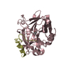
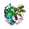
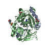
 Components Components | Strictosidine synthase | ||||||
 Keywords Keywords | LYASE / ALKALOID METABOLISM / SIX BLADED BETA PROPELLER FOLD / STR1 / VACUOLE / SYNTHASE / GLYCOPROTEIN | ||||||
| Function / homology |  Function and homology information Function and homology informationstrictosidine synthase / strictosidine synthase activity / indole alkaloid metabolic process / alkaloid biosynthetic process / vacuole / endomembrane system Similarity search - Function | ||||||
| Biological species |  Rauvolfia serpentina (serpentwood) Rauvolfia serpentina (serpentwood) | ||||||
| Method |  X-RAY DIFFRACTION / SYNCHROTRON / MOLECULAR REPLACEMENT / Resolution: 2.549 Å X-RAY DIFFRACTION / SYNCHROTRON / MOLECULAR REPLACEMENT / Resolution: 2.549 Å | ||||||
 Authors Authors | Cai, Y. / Shao, N. / Xie, H. / Futamura, Y. / Panjikar, S. / Liu, H. / Zhu, H. / Osada, H. / Zou, H. | ||||||
| Funding support |  China, 1items China, 1items
| ||||||
 Citation Citation | Journal: to be published Title: Crystal Structure of Strictosidine in complex with 1H-indole-4-ethanamine Authors: Cai, Y. / Shao, N. / Xie, H. / Futamura, Y. / Panjikar, S. / Liu, H. / Zhu, H. / Osada, H. / Zou, H. #1: Journal: Plant Cell / Year: 2006Title: The structure of Rauvolfia serpentina strictosidine synthase is a novel six-bladed beta-propeller fold in plant proteins. Authors: Ma, X. / Panjikar, S. / Koepke, J. / Loris, E. / Stoeckigt, J. | ||||||
| History |
|
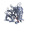
- Structure visualization
| Structure viewer | Molecule: MolmilJmol/JSmol |
|---|
- Downloads & links
-Download
| PDBx/mmCIF format | 6n5v.cif.gz | 132.1 KB | Display | PDBx/mmCIF format |
|---|---|---|---|---|
| PDB format | pdb6n5v.ent.gz | 102.3 KB | Display | PDB format |
| PDBx/mmJSON format | 6n5v.json.gz | Tree view | PDBx/mmJSON format | |
| Others |  Other downloads Other downloads |
-Validation report
| Arichive directory | https://data.pdbj.org/pub/pdb/validation_reports/n5/6n5vftp://data.pdbj.org/pub/pdb/validation_reports/n5/6n5v | HTTPS FTP |
|---|
-Related structure data
| Related structure data |  2fp8C 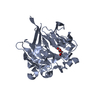 2fp9C 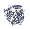 2fpbC  2fpcC  2v91S S: Starting model for refinement C: citing same article ( |
|---|---|
| Similar structure data |


-Links
 PDBj
PDBj


- Assembly
| Deposited unit | 
| ||||||||
|---|---|---|---|---|---|---|---|---|---|
| 1 | 
| ||||||||
| 2 | 
| ||||||||
| Unit cell |
|
-Components
| #1: Protein | Mass: 35207.309 Da / Num. of mol.: 2 Source method: isolated from a genetically manipulated source Source: (gene. exp.) Rauvolfia serpentina (serpentwood) / Gene: STR1 / Plasmid: PQE-2 / Production host:  #2: Chemical | 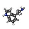  Mass: 160.216 Da / Num. of mol.: 2 / Source method: obtained synthetically / Formula: C10H12N2 / Feature type: SUBJECT OF INVESTIGATION Mass: 160.216 Da / Num. of mol.: 2 / Source method: obtained synthetically / Formula: C10H12N2 / Feature type: SUBJECT OF INVESTIGATION#3: Water | ChemComp-HOH / |  Mass: 18.015 Da / Num. of mol.: 9 / Source method: isolated from a natural source / Formula: H2O Mass: 18.015 Da / Num. of mol.: 9 / Source method: isolated from a natural source / Formula: H2OHas protein modification | Y | |
|---|
-Experimental details
-Experiment
| Experiment | Method: X-RAY DIFFRACTION / Number of used crystals: 1 |
|---|
- Sample preparation
| Crystal | Density Matthews: 3.58 Å3/Da / Density % sol: 68.8 % / Description: Hexagonal |
|---|---|
| Crystal grow | Temperature: 300 K / Method: vapor diffusion / pH: 7.5 Details: 0.8 M potassium sodium tartrate tetrahydrate and 0.1 M HEPES, pH 7.4, 5 mg/mL strictosidine synthase, 3 mM 4-IEA (1H-indole-4-ethanamine) |
-Data collection
| Diffraction | Mean temperature: 100 K / Serial crystal experiment: N |
|---|---|
| Diffraction source | Source: SYNCHROTRON / Site: SSRF / Beamline: BL17U1 / Wavelength: 0.97915 Å |
| Detector | Type: ADSC QUANTUM 210r / Detector: CCD / Date: Jul 1, 2017 / Details: DCM |
| Radiation | Monochromator: SI / Protocol: SINGLE WAVELENGTH / Monochromatic (M) / Laue (L): M / Scattering type: x-ray |
| Radiation wavelength | Wavelength: 0.97915 Å / Relative weight: 1 |
| Reflection | Resolution: 2.549→37.6 Å / Num. obs: 64264 / % possible obs: 95.2 % / Redundancy: 2.23 % / Biso Wilson estimate: 65 Å2 / CC1/2: 0.996 / Rmerge(I) obs: 0.06 / Rrim(I) all: 0.064 / Net I/σ(I): 9.3 |
| Reflection shell | Resolution: 2.55→2.7 Å / Redundancy: 2.27 % / Rmerge(I) obs: 0.71 / Mean I/σ(I) obs: 1.5 / Num. unique obs: 10128 / CC1/2: 0.906 / Rrim(I) all: 0.839 / % possible all: 93.4 |
- Processing
| Software |
| ||||||||||||||||||||||||||||||||||||||||||||||||||||||||
|---|---|---|---|---|---|---|---|---|---|---|---|---|---|---|---|---|---|---|---|---|---|---|---|---|---|---|---|---|---|---|---|---|---|---|---|---|---|---|---|---|---|---|---|---|---|---|---|---|---|---|---|---|---|---|---|---|---|
| Refinement | Method to determine structure: MOLECULAR REPLACEMENT Starting model: 2V91 Resolution: 2.549→37.6 Å / SU ML: 0.4 / Cross valid method: FREE R-VALUE / σ(F): 1.15 / Phase error: 33.89
| ||||||||||||||||||||||||||||||||||||||||||||||||||||||||
| Solvent computation | Shrinkage radii: 0.9 Å / VDW probe radii: 1.11 Å | ||||||||||||||||||||||||||||||||||||||||||||||||||||||||
| Displacement parameters | Biso mean: 81 Å2 | ||||||||||||||||||||||||||||||||||||||||||||||||||||||||
| Refinement step | Cycle: LAST / Resolution: 2.549→37.6 Å
| ||||||||||||||||||||||||||||||||||||||||||||||||||||||||
| Refine LS restraints |
| ||||||||||||||||||||||||||||||||||||||||||||||||||||||||
| LS refinement shell |
|
