 Movie
Movie Controller


+ Open data

- Basic information
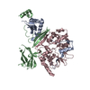
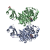
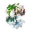
| Entry | Database: PDB / ID: 6zjo | ||||||||||||
|---|---|---|---|---|---|---|---|---|---|---|---|---|---|
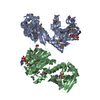
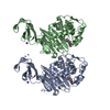
| Title | Crystal Structure of Staphylococcus aureus RsgA. | ||||||||||||
 Components Components | Small ribosomal subunit biogenesis GTPase RsgA | ||||||||||||
 Keywords Keywords | RNA BINDING PROTEIN / GTPase / TRAFAC / ribosome assembly | ||||||||||||
| Function / homology | PHOSPHATE ION / :  Function and homology information Function and homology information | ||||||||||||
| Biological species |   Staphylococcus aureus subsp. aureus (bacteria) Staphylococcus aureus subsp. aureus (bacteria) | ||||||||||||
| Method |  X-RAY DIFFRACTION / SYNCHROTRON / MOLECULAR REPLACEMENT / Resolution: 2.01 Å X-RAY DIFFRACTION / SYNCHROTRON / MOLECULAR REPLACEMENT / Resolution: 2.01 Å | ||||||||||||
 Authors Authors | Bennison, D.J. / Rafferty, J.B. / Corrigan, R.M. | ||||||||||||
| Funding support |  United Kingdom, 3items United Kingdom, 3items
| ||||||||||||
 Citation Citation | Journal: Mbio / Year: 2021 Title: The Stringent Response Inhibits 70S Ribosome Formation in Staphylococcus aureus by Impeding GTPase-Ribosome Interactions. Authors: Bennison, D.J. / Nakamoto, J.A. / Craggs, T.D. / Milon, P. / Rafferty, J.B. / Corrigan, R.M. | ||||||||||||
| History |
|
- Structure visualization
| Structure viewer | Molecule: MolmilJmol/JSmol |
|---|
- Downloads & links
-Download
| PDBx/mmCIF format | 6zjo.cif.gz | 129.5 KB | Display | PDBx/mmCIF format |
|---|---|---|---|---|
| PDB format | pdb6zjo.ent.gz | 97.6 KB | Display | PDB format |
| PDBx/mmJSON format | 6zjo.json.gz | Tree view | PDBx/mmJSON format | |
| Others |  Other downloads Other downloads |
-Validation report
| Arichive directory | https://data.pdbj.org/pub/pdb/validation_reports/zj/6zjoftp://data.pdbj.org/pub/pdb/validation_reports/zj/6zjo | HTTPS FTP |
|---|
-Related structure data
| Related structure data | 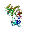 6zhlSC  6zhmC S: Starting model for refinement C: citing same article ( |
|---|---|
| Similar structure data |


-Links
 PDBj
PDBj- Assembly
| Deposited unit | 
| ||||||||
|---|---|---|---|---|---|---|---|---|---|
| 1 |
| ||||||||
| Unit cell |
|
-Components
| #1: Protein | Mass: 36111.777 Da / Num. of mol.: 2 Source method: isolated from a genetically manipulated source Source: (gene. exp.) Staphylococcus aureus subsp. aureus (bacteria)Plasmid: pET28b / Production host: References: UniProt: A0A4V5LHH4, Hydrolases; Acting on acid anhydrides; In phosphorus-containing anhydrides #2: Chemical |   Mass: 65.409 Da / Num. of mol.: 2 / Source method: obtained synthetically / Formula: Zn Mass: 65.409 Da / Num. of mol.: 2 / Source method: obtained synthetically / Formula: Zn#3: Chemical | ChemComp-EDO /   Mass: 62.068 Da / Num. of mol.: 16 / Source method: obtained synthetically / Formula: C2H6O2 Mass: 62.068 Da / Num. of mol.: 16 / Source method: obtained synthetically / Formula: C2H6O2#4: Chemical |   Mass: 94.971 Da / Num. of mol.: 3 / Source method: obtained synthetically / Formula: PO4 Mass: 94.971 Da / Num. of mol.: 3 / Source method: obtained synthetically / Formula: PO4#5: Water | ChemComp-HOH / |  Mass: 18.015 Da / Num. of mol.: 212 / Source method: isolated from a natural source / Formula: H2O Mass: 18.015 Da / Num. of mol.: 212 / Source method: isolated from a natural source / Formula: H2OHas ligand of interest | N | |
|---|
-Experimental details
-Experiment
| Experiment | Method: X-RAY DIFFRACTION / Number of used crystals: 1 |
|---|
- Sample preparation
| Crystal | Density Matthews: 2.71 Å3/Da / Density % sol: 54.61 % / Description: A single rod-shaped crystal |
|---|---|
| Crystal grow | Temperature: 290 K / Method: vapor diffusion, sitting drop / pH: 6 Details: 0.15 M ammonium sulphate, 0.1 M MES, 15% (w/v) PEG 4000 |
-Data collection
| Diffraction | Mean temperature: 100 K / Serial crystal experiment: N | ||||||||||||||||||||||||||||||
|---|---|---|---|---|---|---|---|---|---|---|---|---|---|---|---|---|---|---|---|---|---|---|---|---|---|---|---|---|---|---|---|
| Diffraction source | Source: SYNCHROTRON / Site: Diamond / Beamline: I04 / Wavelength: 0.97949 Å | ||||||||||||||||||||||||||||||
| Detector | Type: DECTRIS EIGER2 X 16M / Detector: PIXEL / Date: May 13, 2018 | ||||||||||||||||||||||||||||||
| Radiation | Monochromator: M / Protocol: SINGLE WAVELENGTH / Monochromatic (M) / Laue (L): M / Scattering type: x-ray | ||||||||||||||||||||||||||||||
| Radiation wavelength | Wavelength: 0.97949 Å / Relative weight: 1 | ||||||||||||||||||||||||||||||
| Reflection | Resolution: 2.01→47.2 Å / Num. obs: 44789 / % possible obs: 98.2 % / Redundancy: 6.8 % / CC1/2: 0.997 / Rmerge(I) obs: 0.137 / Rpim(I) all: 0.057 / Rrim(I) all: 0.149 / Net I/σ(I): 8.4 / Num. measured all: 306023 / Scaling rejects: 195 | ||||||||||||||||||||||||||||||
| Reflection shell | Diffraction-ID: 1
|
- Processing
| Software |
| ||||||||||||||||||
|---|---|---|---|---|---|---|---|---|---|---|---|---|---|---|---|---|---|---|---|
| Refinement | Method to determine structure: MOLECULAR REPLACEMENT Starting model: 6ZHL Resolution: 2.01→47.196 Å / Cross valid method: THROUGHOUT / σ(F): 0 Details: HYDROGENS HAVE BEEN ADDED IN THE RIDING POSITIONS U VALUES :REFINED INDIVIDUALLY
| ||||||||||||||||||
| Displacement parameters | Biso max: 123.45 Å2 / Biso mean: 40.2119 Å2 / Biso min: 17.89 Å2 | ||||||||||||||||||
| Refinement step | Cycle: LAST / Resolution: 2.01→47.196 Å
|
