 Movie
Movie Controller

[English] 日本語
 Yorodumi
- PDB-6zgx: Structure of human galactokinase 1 bound with 2-(4-chlorophenyl)-... -
+ Open data

- Basic information
| Entry | Database: PDB / ID: 6zgx | ||||||
|---|---|---|---|---|---|---|---|
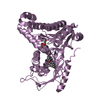
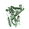
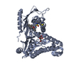
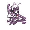
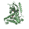
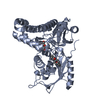
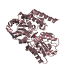
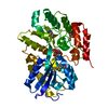
| Title | Structure of human galactokinase 1 bound with 2-(4-chlorophenyl)-N-(pyrimidin-2-yl)acetamide | ||||||
 Components Components | Galactokinase | ||||||
 Keywords Keywords | TRANSFERASE / GALK1 / galactokinase 1 / fragment screening / allosteric fragment / binding hotspt | ||||||
| Function / homology |  Function and homology information Function and homology informationglycolytic process from galactose / Defective GALK1 causes GALCT2 / galactitol metabolic process / galactokinase / galactokinase activity / beta-D-galactose catabolic process via UDP-galactose, Leloir pathway / galactose binding / galactose metabolic process / Galactose catabolism / extracellular exosome ...glycolytic process from galactose / Defective GALK1 causes GALCT2 / galactitol metabolic process / galactokinase / galactokinase activity / beta-D-galactose catabolic process via UDP-galactose, Leloir pathway / galactose binding / galactose metabolic process / Galactose catabolism / extracellular exosome / ATP binding / membrane / cytoplasm / cytosol Similarity search - Function | ||||||
| Biological species |  Homo sapiens (human) Homo sapiens (human) | ||||||
| Method |  X-RAY DIFFRACTION / SYNCHROTRON / MOLECULAR REPLACEMENT / molecular replacement / Resolution: 1.86 Å X-RAY DIFFRACTION / SYNCHROTRON / MOLECULAR REPLACEMENT / molecular replacement / Resolution: 1.86 Å | ||||||
 Authors Authors | Mackinnon, S.R. / Bezerra, G.A. / Zhang, M. / Foster, W. / Krojer, T. / Brandao-Neto, J. / Douangamath, A. / Arrowsmith, C. / Edwards, A. / Bountra, C. ...Mackinnon, S.R. / Bezerra, G.A. / Zhang, M. / Foster, W. / Krojer, T. / Brandao-Neto, J. / Douangamath, A. / Arrowsmith, C. / Edwards, A. / Bountra, C. / Brennan, P. / Lai, K. / Yue, W.W. | ||||||
| Funding support |  United Kingdom, 1items United Kingdom, 1items
| ||||||
 Citation Citation | Journal: Acs Chem.Biol. / Year: 2021 Title: Fragment Screening Reveals Starting Points for Rational Design of Galactokinase 1 Inhibitors to Treat Classic Galactosemia. Authors: Mackinnon, S.R. / Krojer, T. / Foster, W.R. / Diaz-Saez, L. / Tang, M. / Huber, K.V.M. / von Delft, F. / Lai, K. / Brennan, P.E. / Arruda Bezerra, G. / Yue, W.W. | ||||||
| History |
|
- Structure visualization
| Structure viewer | Molecule: MolmilJmol/JSmol |
|---|
- Downloads & links
-Download
| PDBx/mmCIF format | 6zgx.cif.gz | 299.1 KB | Display | PDBx/mmCIF format |
|---|---|---|---|---|
| PDB format | pdb6zgx.ent.gz | 236.6 KB | Display | PDB format |
| PDBx/mmJSON format | 6zgx.json.gz | Tree view | PDBx/mmJSON format | |
| Others |  Other downloads Other downloads |
-Validation report
| Arichive directory | https://data.pdbj.org/pub/pdb/validation_reports/zg/6zgxftp://data.pdbj.org/pub/pdb/validation_reports/zg/6zgx | HTTPS FTP |
|---|
-Related structure data
| Related structure data | 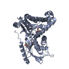 6q3xC 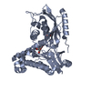 6zfhC 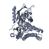 6zgvC 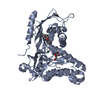 6zgwC  6zgyC  6zgzC  6zh0C 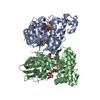 1wuuS S: Starting model for refinement C: citing same article ( |
|---|---|
| Similar structure data |


-Links
 PDBj
PDBj

- Assembly
| Deposited unit | 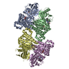
| ||||||||
|---|---|---|---|---|---|---|---|---|---|
| 1 | 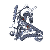
| ||||||||
| 2 | 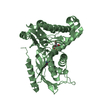
| ||||||||
| 3 | 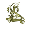
| ||||||||
| 4 | 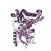
| ||||||||
| Unit cell |
|
-Components
| #1: Protein | Mass: 43102.977 Da / Num. of mol.: 4 / Mutation: K252A, E253A Source method: isolated from a genetically manipulated source Source: (gene. exp.) Homo sapiens (human) / Gene: GALK1, GALK / Production host:  #2: Sugar |   Type: D-saccharide, beta linking / Mass: 180.156 Da / Num. of mol.: 3 Type: D-saccharide, beta linking / Mass: 180.156 Da / Num. of mol.: 3Source method: isolated from a genetically manipulated source Formula: C6H12O6 #3: Chemical |   Mass: 350.414 Da / Num. of mol.: 3 / Source method: obtained synthetically / Formula: C20H22N4O2 Mass: 350.414 Da / Num. of mol.: 3 / Source method: obtained synthetically / Formula: C20H22N4O2#4: Chemical | ChemComp-S6V / |   Mass: 247.293 Da / Num. of mol.: 1 / Source method: obtained synthetically / Formula: C13H17N3O2 / Feature type: SUBJECT OF INVESTIGATION Mass: 247.293 Da / Num. of mol.: 1 / Source method: obtained synthetically / Formula: C13H17N3O2 / Feature type: SUBJECT OF INVESTIGATION#5: Water | ChemComp-HOH / |  Mass: 18.015 Da / Num. of mol.: 456 / Source method: isolated from a natural source / Formula: H2O Mass: 18.015 Da / Num. of mol.: 456 / Source method: isolated from a natural source / Formula: H2OHas ligand of interest | Y | Has protein modification | Y | |
|---|
-Experimental details
-Experiment
| Experiment | Method: X-RAY DIFFRACTION / Number of used crystals: 1 |
|---|
- Sample preparation
| Crystal | Density Matthews: 2.97 Å3/Da / Density % sol: 58.62 % |
|---|---|
| Crystal grow | Temperature: 293 K / Method: vapor diffusion, sitting drop Details: 0.1 M MOPS/sodium HEPES pH 7.0-7.5, 40-50 % Morpheus Precipitant Mix 4 (50% mix = 12.5% MPD, 12.5% PEG1000, 12.5% PEG3350), 0.1 M Morpheus Carboxylic acids mix (0.02M each of - sodium ...Details: 0.1 M MOPS/sodium HEPES pH 7.0-7.5, 40-50 % Morpheus Precipitant Mix 4 (50% mix = 12.5% MPD, 12.5% PEG1000, 12.5% PEG3350), 0.1 M Morpheus Carboxylic acids mix (0.02M each of - sodium formate, ammonium acetate, sodium citrate tribasic dehydrate, sodium potassium tartrate tetrahydrate and sodium oxamate) PH range: 7.0-7.5 |
-Data collection
| Diffraction | Mean temperature: 100 K / Serial crystal experiment: N | |||||||||||||||||||||||||||
|---|---|---|---|---|---|---|---|---|---|---|---|---|---|---|---|---|---|---|---|---|---|---|---|---|---|---|---|---|
| Diffraction source | Source: SYNCHROTRON / Site: Diamond / Beamline: I04-1 / Wavelength: 0.91587 Å | |||||||||||||||||||||||||||
| Detector | Type: DECTRIS PILATUS 6M / Detector: PIXEL / Date: Oct 1, 2018 | |||||||||||||||||||||||||||
| Radiation | Protocol: SINGLE WAVELENGTH / Monochromatic (M) / Laue (L): M / Scattering type: x-ray | |||||||||||||||||||||||||||
| Radiation wavelength | Wavelength: 0.91587 Å / Relative weight: 1 | |||||||||||||||||||||||||||
| Reflection | Resolution: 1.86→114.44 Å / Num. obs: 164750 / % possible obs: 99.6 % / Redundancy: 3.4 % / CC1/2: 0.988 / Rmerge(I) obs: 0.081 / Rpim(I) all: 0.052 / Rrim(I) all: 0.096 / Net I/σ(I): 7 / Num. measured all: 560340 / Scaling rejects: 264 | |||||||||||||||||||||||||||
| Reflection shell | Diffraction-ID: 1 / Redundancy: 3.5 %
|
-Phasing
| Phasing | Method: molecular replacement |
|---|
- Processing
| Software |
| |||||||||||||||||||||||||||||||||||||||||||||
|---|---|---|---|---|---|---|---|---|---|---|---|---|---|---|---|---|---|---|---|---|---|---|---|---|---|---|---|---|---|---|---|---|---|---|---|---|---|---|---|---|---|---|---|---|---|---|
| Refinement | Method to determine structure: MOLECULAR REPLACEMENT Starting model: 1WUU Resolution: 1.86→82.59 Å / Cor.coef. Fo:Fc: 0.94 / Cor.coef. Fo:Fc free: 0.929 / SU B: 5.844 / SU ML: 0.159 / Cross valid method: THROUGHOUT / σ(F): 0 / ESU R: 0.153 / ESU R Free: 0.141 / Stereochemistry target values: MAXIMUM LIKELIHOOD / Details: U VALUES : REFINED INDIVIDUALLY
| |||||||||||||||||||||||||||||||||||||||||||||
| Solvent computation | Ion probe radii: 0.8 Å / Shrinkage radii: 0.8 Å / VDW probe radii: 1.2 Å / Solvent model: MASK | |||||||||||||||||||||||||||||||||||||||||||||
| Displacement parameters | Biso max: 120.86 Å2 / Biso mean: 38.869 Å2 / Biso min: 15.22 Å2
| |||||||||||||||||||||||||||||||||||||||||||||
| Refinement step | Cycle: final / Resolution: 1.86→82.59 Å
| |||||||||||||||||||||||||||||||||||||||||||||
| Refine LS restraints |
| |||||||||||||||||||||||||||||||||||||||||||||
| LS refinement shell | Resolution: 1.86→1.907 Å / Rfactor Rfree error: 0
|
