Biotechnology and Biological Sciences Research Council (BBSRC)
BB/S003339/1
英国
Wellcome Trust
202933/Z/16/Z
英国
引用
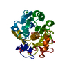
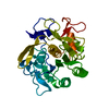
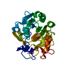
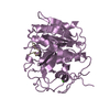
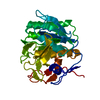
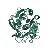
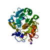
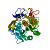
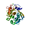
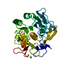
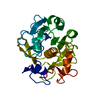
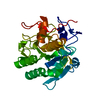
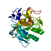
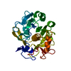
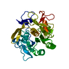
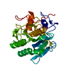
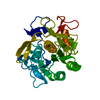
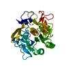
ジャーナル: Front Mol Biosci / 年: 2020 タイトル: A Workflow for Protein Structure Determination From Thin Crystal Lamella by Micro-Electron Diffraction. 著者: Emma V Beale / David G Waterman / Corey Hecksel / Jason van Rooyen / James B Gilchrist / James M Parkhurst / Felix de Haas / Bart Buijsse / Gwyndaf Evans / Peijun Zhang / 要旨: MicroED has recently emerged as a powerful method for the analysis of biological structures at atomic resolution. This technique has been largely limited to protein nanocrystals which grow either as ...MicroED has recently emerged as a powerful method for the analysis of biological structures at atomic resolution. This technique has been largely limited to protein nanocrystals which grow either as needles or plates measuring only a few hundred nanometers in thickness. Furthermore, traditional microED data processing uses established X-ray crystallography software that is not optimized for handling compound effects that are unique to electron diffraction data. Here, we present an integrated workflow for microED, from sample preparation by cryo-focused ion beam milling, through data collection with a standard Ceta-D detector, to data processing using the DIALS software suite, thus enabling routine atomic structure determination of protein crystals of any size and shape using microED. We demonstrate the effectiveness of the workflow by determining the structure of proteinase K to 2.0 Å resolution and show the advantage of using protein crystal lamellae over nanocrystals.
Imaging-ID: 1 / 平均露光時間: 0.85 sec. / 電子線照射量: 0.034 e/Å2 / フィルム・検出器のモデル: OTHER / 撮影したグリッド数: 1 / 詳細: The images were recorded using a Ceta-D detector operated in rolling-shutter mode with 2x2 binning.
 ムービー
ムービー コントローラー


 データを開く
 基本情報
基本情報 要素
要素 キーワード
キーワード 機能・相同性情報
 Parengyodontium album (菌類)
 分子置換 / クライオ電子顕微鏡法 / 解像度: 2.4 Å
  データ登録者
 英国, 3件
  引用
引用

 構造の表示
構造の表示 ダウンロードとリンク
 その他のダウンロード


 PDBj
PDBj

 集合体
集合体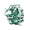

 分子量: 40.078 Da / 分子数: 1 / 由来タイプ: 合成 / 式: Ca
 分子量: 18.015 Da / 分子数: 6 / 由来タイプ: 天然 / 式: H2O
 試料調製
試料調製
 解析
解析