Movie
Movie Controller

[English] 日本語
 Yorodumi
- PDB-6z7p: Composite model of the Caulobacter crescentus S-layer bound to th... -
+ Open data

- Basic information
| Entry | Database: PDB / ID: 6z7p | |||||||||
|---|---|---|---|---|---|---|---|---|---|---|
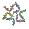
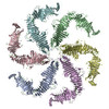
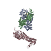
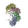
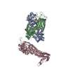
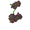
| Title | Composite model of the Caulobacter crescentus S-layer bound to the O-antigen of lipopolysaccharide | |||||||||
 Components Components | S-layer protein | |||||||||
 Keywords Keywords | STRUCTURAL PROTEIN / RsaA S-layer sub-tomogram averaging Caulobacter lipopolysaccharide O-antigen | |||||||||
| Function / homology | RsaA N-terminal domain / S-layer / RTX calcium-binding nonapeptide repeat / RTX calcium-binding nonapeptide repeat (4 copies) / Serralysin-like metalloprotease, C-terminal / calcium ion binding / S-layer protein Function and homology information Function and homology information | |||||||||
| Biological species |  Caulobacter vibrioides (bacteria) Caulobacter vibrioides (bacteria) | |||||||||
| Method | ELECTRON MICROSCOPY / subtomogram averaging / cryo EM / Resolution: 4.8 Å | |||||||||
 Authors Authors | Bharat, T.A.M. / von Kugelgen, A. | |||||||||
| Funding support |  United Kingdom, 1items United Kingdom, 1items
| |||||||||
 Citation Citation | Journal: Cell / Year: 2020 Title: In Situ Structure of an Intact Lipopolysaccharide-Bound Bacterial Surface Layer. Authors: Andriko von Kügelgen / Haiping Tang / Gail G Hardy / Danguole Kureisaite-Ciziene / Yves V Brun / Phillip J Stansfeld / Carol V Robinson / Tanmay A M Bharat /   Abstract: Most bacterial and all archaeal cells are encapsulated by a paracrystalline, protective, and cell-shape-determining proteinaceous surface layer (S-layer). On Gram-negative bacteria, S-layers are ...Most bacterial and all archaeal cells are encapsulated by a paracrystalline, protective, and cell-shape-determining proteinaceous surface layer (S-layer). On Gram-negative bacteria, S-layers are anchored to cells via lipopolysaccharide. Here, we report an electron cryomicroscopy structure of the Caulobacter crescentus S-layer bound to the O-antigen of lipopolysaccharide. Using native mass spectrometry and molecular dynamics simulations, we deduce the length of the O-antigen on cells and show how lipopolysaccharide binding and S-layer assembly is regulated by calcium. Finally, we present a near-atomic resolution in situ structure of the complete S-layer using cellular electron cryotomography, showing S-layer arrangement at the tip of the O-antigen. A complete atomic structure of the S-layer shows the power of cellular tomography for in situ structural biology and sheds light on a very abundant class of self-assembling molecules with important roles in prokaryotic physiology with marked potential for synthetic biology and surface-display applications. #1: Journal: Nat Microbiol / Year: 2017Title: Structure of the hexagonal surface layer on Caulobacter crescentus cells. Authors: Tanmay A M Bharat / Danguole Kureisaite-Ciziene / Gail G Hardy / Ellen W Yu / Jessica M Devant / Wim J H Hagen / Yves V Brun / John A G Briggs / Jan Löwe /  Abstract: Many prokaryotic cells are encapsulated by a surface layer (S-layer) consisting of repeating units of S-layer proteins. S-layer proteins are a diverse class of molecules found in Gram-positive and ...Many prokaryotic cells are encapsulated by a surface layer (S-layer) consisting of repeating units of S-layer proteins. S-layer proteins are a diverse class of molecules found in Gram-positive and Gram-negative bacteria and most archaea. S-layers protect cells from the outside, provide mechanical stability and also play roles in pathogenicity. In situ structural information about this highly abundant class of proteins is scarce, so atomic details of how S-layers are arranged on the surface of cells have remained elusive. Here, using purified Caulobacter crescentus' sole S-layer protein RsaA, we obtained a 2.7 Å X-ray structure that shows the hexameric S-layer lattice. We also solved a 7.4 Å structure of the S-layer through electron cryotomography and sub-tomogram averaging of cell stalks. The X-ray structure was docked unambiguously into the electron cryotomography map, resulting in a pseudo-atomic-level description of the in vivo S-layer, which agrees completely with the atomic X-ray lattice model. The cellular S-layer atomic structure shows that the S-layer is porous, with a largest gap dimension of 27 Å, and is stabilized by multiple Ca ions bound near the interfaces. This study spans different spatial scales from atoms to cells by combining X-ray crystallography with electron cryotomography and sub-nanometre-resolution sub-tomogram averaging. | |||||||||
| History |
|
- Structure visualization
| Movie |
Movie viewer |
|---|---|
| Structure viewer | Molecule: MolmilJmol/JSmol |
- Downloads & links
-Download
| PDBx/mmCIF format | 6z7p.cif.gz | 173.1 KB | Display | PDBx/mmCIF format |
|---|---|---|---|---|
| PDB format | pdb6z7p.ent.gz | 126.8 KB | Display | PDB format |
| PDBx/mmJSON format | 6z7p.json.gz | Tree view | PDBx/mmJSON format | |
| Others |  Other downloads Other downloads |
-Validation report
| Arichive directory | https://data.pdbj.org/pub/pdb/validation_reports/z7/6z7pftp://data.pdbj.org/pub/pdb/validation_reports/z7/6z7p | HTTPS FTP |
|---|
-Related structure data
| Related structure data | 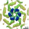 10388MC  6t72C M: map data used to model this data C: citing same article ( |
|---|---|
| Similar structure data |


-Links
 PDBj
PDBj- Assembly
| Deposited unit | 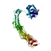
|
|---|---|
| 1 |
|
-Components
| #1: Protein | Mass: 98022.703 Da / Num. of mol.: 1 / Source method: isolated from a natural source Source: (natural) Caulobacter vibrioides (strain ATCC 19089 / CB15) (bacteria)Strain: ATCC 19089 / CB15 / References: UniProt: P35828 | ||
|---|---|---|---|
| #2: Polysaccharide | 4-acetamido-4,6-dideoxy-alpha-D-mannopyranose-(1-3)-4-acetamido-4,6-dideoxy-alpha-D-mannopyranose- ...4-acetamido-4,6-dideoxy-alpha-D-mannopyranose-(1-3)-4-acetamido-4,6-dideoxy-alpha-D-mannopyranose-(1-3)-beta-D-mannopyranose-(1-3)-4-acetamido-4,6-dideoxy-alpha-D-mannopyranose-(1-3)-4-acetamido-4,6-dideoxy-alpha-D-mannopyranose-(1-3)-beta-D-mannopyranose-(1-3)-4-acetamido-4,6-dideoxy-alpha-D-mannopyranose-(1-3)-4-acetamido-4,6-dideoxy-alpha-D-mannopyranose-(1-3)-beta-D-mannopyranose | ||
| #3: Chemical | ChemComp-CA /   Mass: 40.078 Da / Num. of mol.: 22 / Source method: obtained synthetically / Formula: Ca / Feature type: SUBJECT OF INVESTIGATION Mass: 40.078 Da / Num. of mol.: 22 / Source method: obtained synthetically / Formula: Ca / Feature type: SUBJECT OF INVESTIGATIONHas ligand of interest | Y | |
-Experimental details
-Experiment
| Experiment | Method: ELECTRON MICROSCOPY |
|---|---|
| EM experiment | Aggregation state: CELL / 3D reconstruction method: subtomogram averaging |
- Sample preparation
| Component | Name: S-layer / Type: COMPLEX / Entity ID: #1 / Source: NATURAL |
|---|---|
| Source (natural) | Organism: Caulobacter vibrioides NA1000 (bacteria) / Strain: YB2811 / Cellular location: extra-cellular |
| Buffer solution | pH: 7 / Details: PYE |
| Specimen | Embedding applied: NO / Shadowing applied: NO / Staining applied: NO / Vitrification applied: YES / Details: Caulobacter crescentus stalk |
| Specimen support | Details: 15 mA / Grid material: COPPER/RHODIUM / Grid mesh size: 200 divisions/in. / Grid type: Quantifoil R2/2 |
| Vitrification | Instrument: FEI VITROBOT MARK IV / Cryogen name: NITROGEN / Humidity: 100 % / Chamber temperature: 283.15 K / Details: 1.5 s blot |
- Electron microscopy imaging
| Experimental equipment |  Model: Titan Krios / Image courtesy: FEI Company |
|---|---|
| Microscopy | Model: FEI TITAN KRIOS |
| Electron gun | Electron source:  FIELD EMISSION GUN / Accelerating voltage: 300 kV / Illumination mode: FLOOD BEAM FIELD EMISSION GUN / Accelerating voltage: 300 kV / Illumination mode: FLOOD BEAM |
| Electron lens | Mode: BRIGHT FIELD / Nominal magnification: 105000 X / Calibrated magnification: 105000 X / Nominal defocus max: -5000 nm / Nominal defocus min: -2000 nm / Calibrated defocus min: -2000 nm / Calibrated defocus max: -5000 nm / Cs: 2.7 mm / C2 aperture diameter: 70 µm / Alignment procedure: COMA FREE |
| Specimen holder | Cryogen: NITROGEN / Specimen holder model: FEI TITAN KRIOS AUTOGRID HOLDER / Temperature (max): 70 K / Temperature (min): 70 K |
| Image recording | Average exposure time: 1 sec. / Electron dose: 3.4 e/Å2 / Detector mode: SUPER-RESOLUTION / Film or detector model: GATAN K2 QUANTUM (4k x 4k) / Num. of grids imaged: 1 / Details: Dose symmetric tilt scheme (Hagen et al, JSB) |
| EM imaging optics | Energyfilter name: GIF Quantum LS / Chromatic aberration corrector: not used / Energyfilter slit width: 20 eV / Spherical aberration corrector: not used |
- Processing
| EM software |
| |||||||||||||||||||||||||
|---|---|---|---|---|---|---|---|---|---|---|---|---|---|---|---|---|---|---|---|---|---|---|---|---|---|---|
| CTF correction | Details: Following Turonova and Briggs NovaCTF / Type: PHASE FLIPPING AND AMPLITUDE CORRECTION | |||||||||||||||||||||||||
| Symmetry | Point symmetry: C1 (asymmetric) | |||||||||||||||||||||||||
| 3D reconstruction | Resolution: 4.8 Å / Resolution method: FSC 0.143 CUT-OFF / Num. of particles: 51866 / Algorithm: FOURIER SPACE Details: Tomogram generation in IMOD using the NovaCTF implementation of Turonova and Briggs. Sub-tomogram averaging performed using the AV3 package (Friedrich Foerster) applied to tubular specimens ...Details: Tomogram generation in IMOD using the NovaCTF implementation of Turonova and Briggs. Sub-tomogram averaging performed using the AV3 package (Friedrich Foerster) applied to tubular specimens (Bharat et al, PLoS Biology, 2011) Num. of class averages: 1 / Symmetry type: POINT | |||||||||||||||||||||||||
| EM volume selection | Method: RELION / Details: RELION subtomogram averaging / Num. of tomograms: 110 / Num. of volumes extracted: 51866 / Reference model: Ab initio | |||||||||||||||||||||||||
| Atomic model building | Protocol: RIGID BODY FIT / Space: REAL / Target criteria: USCF Chimera Details: Initial rigid body fit of the C-terminal X-ray structure (PDB: 5N8P, amino acids 249-1046) and the N-terminal cryo-EM structure (PDB: 6T72, amino acids 2-243) was performed in USCF Chimera. ...Details: Initial rigid body fit of the C-terminal X-ray structure (PDB: 5N8P, amino acids 249-1046) and the N-terminal cryo-EM structure (PDB: 6T72, amino acids 2-243) was performed in USCF Chimera. The missing amino acid linker was manually added suing Coot. The model was not refined against the map and all B-factors were set to zero due to resolution anisotropy. | |||||||||||||||||||||||||
| Atomic model building | 3D fitting-ID: 1 / Pdb chain-ID: A / Source name: PDB / Type: experimental model
|