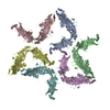
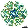
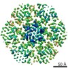
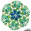
Journal: Nat Microbiol / Year: 2017 Title: Structure of the hexagonal surface layer on Caulobacter crescentus cells. Authors: Tanmay A M Bharat / Danguole Kureisaite-Ciziene / Gail G Hardy / Ellen W Yu / Jessica M Devant / Wim J H Hagen / Yves V Brun / John A G Briggs / Jan Löwe / Abstract: Many prokaryotic cells are encapsulated by a surface layer (S-layer) consisting of repeating units of S-layer proteins. S-layer proteins are a diverse class of molecules found in Gram-positive and ...Many prokaryotic cells are encapsulated by a surface layer (S-layer) consisting of repeating units of S-layer proteins. S-layer proteins are a diverse class of molecules found in Gram-positive and Gram-negative bacteria and most archaea. S-layers protect cells from the outside, provide mechanical stability and also play roles in pathogenicity. In situ structural information about this highly abundant class of proteins is scarce, so atomic details of how S-layers are arranged on the surface of cells have remained elusive. Here, using purified Caulobacter crescentus' sole S-layer protein RsaA, we obtained a 2.7 Å X-ray structure that shows the hexameric S-layer lattice. We also solved a 7.4 Å structure of the S-layer through electron cryotomography and sub-tomogram averaging of cell stalks. The X-ray structure was docked unambiguously into the electron cryotomography map, resulting in a pseudo-atomic-level description of the in vivo S-layer, which agrees completely with the atomic X-ray lattice model. The cellular S-layer atomic structure shows that the S-layer is porous, with a largest gap dimension of 27 Å, and is stabilized by multiple Ca ions bound near the interfaces. This study spans different spatial scales from atoms to cells by combining X-ray crystallography with electron cryotomography and sub-nanometre-resolution sub-tomogram averaging.
A: S-layer protein rsaA B: S-layer protein rsaA C: S-layer protein rsaA D: S-layer protein rsaA E: S-layer protein rsaA F: S-layer protein rsaA hetero molecules
Cryogen: NITROGEN / Specimen holder model: FEI TITAN KRIOS AUTOGRID HOLDER / Temperature (max): 70 K / Temperature (min): 70 K
Image recording
Average exposure time: 1 sec. / Electron dose: 3.4 e/Å2 / Detector mode: SUPER-RESOLUTION / Film or detector model: GATAN K2 SUMMIT (4k x 4k) / Num. of grids imaged: 1 / Details: Dose symmetric tilt scheme (Hagen et al)
EM imaging optics
Energyfilter name: GIF Quantum / Energyfilter upper: 20 eV / Energyfilter lower: 0 eV
-
Processing
EM software
ID
Name
Version
Category
1
RELION
1.4
volumeselection
2
SerialEM
imageacquisition
7
UCSF Chimera
modelfitting
Crystal symmetry
∠γ: 1 ° / C sampling length: 10 Å / A: 220 Å / B: 220 Å / C: 220 Å / Space group name H-M: P121
CTF correction
Details: Following Schur et al, 2016 / Type: PHASE FLIPPING AND AMPLITUDE CORRECTION
3D reconstruction
Resolution: 7.4 Å / Resolution method: FSC 0.143 CUT-OFF / Num. of particles: 51866 / Algorithm: FOURIER SPACE / Num. of class averages: 1 / Symmetry type: 2D CRYSTAL
EM volume selection
Method: RELION / Details: RELION subtomogram averaging / Num. of tomograms: 110 / Num. of volumes extracted: 51866 / Reference model: Ab initio
Atomic model building
Protocol: RIGID BODY FIT / Space: REAL / Target criteria: Cross-correlation coefficient
+
About Yorodumi
-
News
-
Feb 9, 2022. New format data for meta-information of EMDB entries
New format data for meta-information of EMDB entries
Version 3 of the EMDB header file is now the official format.
The previous official version 1.9 will be removed from the archive.
In the structure databanks used in Yorodumi, some data are registered as the other names, "COVID-19 virus" and "2019-nCoV". Here are the details of the virus and the list of structure data.
Jan 31, 2019. EMDB accession codes are about to change! (news from PDBe EMDB page)
EMDB accession codes are about to change! (news from PDBe EMDB page)
The allocation of 4 digits for EMDB accession codes will soon come to an end. Whilst these codes will remain in use, new EMDB accession codes will include an additional digit and will expand incrementally as the available range of codes is exhausted. The current 4-digit format prefixed with “EMD-” (i.e. EMD-XXXX) will advance to a 5-digit format (i.e. EMD-XXXXX), and so on. It is currently estimated that the 4-digit codes will be depleted around Spring 2019, at which point the 5-digit format will come into force.
The EM Navigator/Yorodumi systems omit the EMD- prefix.
Related info.:Q: What is EMD? / ID/Accession-code notation in Yorodumi/EM Navigator
Yorodumi is a browser for structure data from EMDB, PDB, SASBDB, etc.
This page is also the successor to EM Navigator detail page, and also detail information page/front-end page for Omokage search.
The word "yorodu" (or yorozu) is an old Japanese word meaning "ten thousand". "mi" (miru) is to see.
Related info.:EMDB / PDB / SASBDB / Comparison of 3 databanks / Yorodumi Search / Aug 31, 2016. New EM Navigator & Yorodumi / Yorodumi Papers / Jmol/JSmol / Function and homology information / Changes in new EM Navigator and Yorodumi
 Movie
Movie Controller


 Open data
 Basic information
 Components
 Keywords
 Function and homology information
 Caulobacter crescentus NA1000 (bacteria)
 Authors
 United Kingdom,
  Germany, 3items
  Citation
 Structure visualization
 Downloads & links
 Other downloads


 PDBj
PDBj Assembly

 Mass: 40.078 Da / Num. of mol.: 114 / Source method: obtained synthetically / Formula: Ca
 Sample preparation
 Electron microscopy imaging
 FIELD EMISSION GUN / Accelerating voltage: 300 kV / Illumination mode: FLOOD BEAM
 Processing