[English] 日本語
Yorodumi
- PDB-5n8p: S-layer protein RsaA from C. crescentus -

+
Open data


ID or keywords:

Loading...

-
Basic information

Entry
Database: PDB / ID: 5n8p
TitleS-layer protein RsaA from C. crescentus
ComponentsS-layer protein
KeywordsMEMBRANE PROTEIN / S-layer / surface protein
Function / homologyRsaA N-terminal domain / S-layer / RTX calcium-binding nonapeptide repeat / RTX calcium-binding nonapeptide repeat (4 copies) / Serralysin-like metalloprotease, C-terminal / calcium ion binding / extracellular region / S-layer protein
Function and homology information
Biological speciesCaulobacter crescentus CB15 (bacteria)
MethodX-RAY DIFFRACTION / SYNCHROTRON / Resolution: 2.7 Å
AuthorsBharat, T.A.M. / Kureisaite-Ciziene, D. / Lowe, J.
Citation
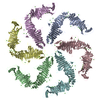
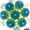
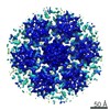
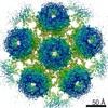
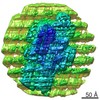
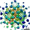
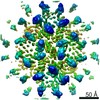
Journal: Nat Microbiol / Year: 2017
Title: Structure of the hexagonal surface layer on Caulobacter crescentus cells.
Authors: Tanmay A M Bharat / Danguole Kureisaite-Ciziene / Gail G Hardy / Ellen W Yu / Jessica M Devant / Wim J H Hagen / Yves V Brun / John A G Briggs / Jan Löwe /
Abstract: Many prokaryotic cells are encapsulated by a surface layer (S-layer) consisting of repeating units of S-layer proteins. S-layer proteins are a diverse class of molecules found in Gram-positive and ...Many prokaryotic cells are encapsulated by a surface layer (S-layer) consisting of repeating units of S-layer proteins. S-layer proteins are a diverse class of molecules found in Gram-positive and Gram-negative bacteria and most archaea. S-layers protect cells from the outside, provide mechanical stability and also play roles in pathogenicity. In situ structural information about this highly abundant class of proteins is scarce, so atomic details of how S-layers are arranged on the surface of cells have remained elusive. Here, using purified Caulobacter crescentus' sole S-layer protein RsaA, we obtained a 2.7 Å X-ray structure that shows the hexameric S-layer lattice. We also solved a 7.4 Å structure of the S-layer through electron cryotomography and sub-tomogram averaging of cell stalks. The X-ray structure was docked unambiguously into the electron cryotomography map, resulting in a pseudo-atomic-level description of the in vivo S-layer, which agrees completely with the atomic X-ray lattice model. The cellular S-layer atomic structure shows that the S-layer is porous, with a largest gap dimension of 27 Å, and is stabilized by multiple Ca ions bound near the interfaces. This study spans different spatial scales from atoms to cells by combining X-ray crystallography with electron cryotomography and sub-nanometre-resolution sub-tomogram averaging.
History
DepositionFeb 24, 2017Deposition site: PDBE / Processing site: PDBE
Revision 1.0Apr 12, 2017Provider: repository / Type: Initial release
Revision 1.1Apr 26, 2017Group: Database references
Revision 1.2Feb 20, 2019Group: Advisory / Data collection / Derived calculations
Category: diffrn_source / pdbx_data_processing_status ...diffrn_source / pdbx_data_processing_status / pdbx_validate_symm_contact / struct_conn / struct_conn_type
Item: _diffrn_source.pdbx_synchrotron_site
Revision 1.3Jul 10, 2019Group: Data collection / Category: diffrn_source / Item: _diffrn_source.pdbx_synchrotron_site

-
Structure visualization

Structure viewerMolecule:
MolmilJmol/JSmol

Downloads & links

-
Assembly

Deposited unit
A: S-layer protein
B: S-layer protein
C: S-layer protein
D: S-layer protein
E: S-layer protein
F: S-layer protein
hetero molecules


Theoretical massNumber of molelcules
Total (without water)593,492120
Polymers588,9236
Non-polymers4,569114
Water2,648147
1


  • Idetical with deposited unit
  • defined by author&software
  • Evidence: microscopy, RsaA polymerises to form a 2D lattice on cells and also in the crystals. This was determined by comparing the crystal packing described here with electron tomography data from ...Evidence: microscopy, RsaA polymerises to form a 2D lattice on cells and also in the crystals. This was determined by comparing the crystal packing described here with electron tomography data from cells (submitted in separate EMDB entry, to be submitted).
TypeNameSymmetry operationNumber
identity operation1_555x,y,z1
Buried area10150 Å2
ΔGint-714 kcal/mol
Surface area168930 Å2
MethodPISA
Unit cell
Length a, b, c (Å)215.880, 74.960, 221.660
Angle α, β, γ (deg.)90.00, 118.99, 90.00
Int Tables number4
Space group name H-MP1211
Noncrystallographic symmetry (NCS)NCS domain:
IDEns-IDDetails
11A
21B
12A
22C
13A
23D
14A
24E
15A
25F
16B
26C
17B
27D
18B
28E
19B
29F
110C
210D
111C
211E
112C
212F
113D
213E
114D
214F
115E
215F

NCS domain segments:
Dom-IDComponent-IDEns-IDRefine codeAuth asym-IDAuth seq-ID
1010A249 - 1026
2010B249 - 1026
1020A249 - 1026
2020C249 - 1026
1030A249 - 1026
2030D249 - 1026
1040A249 - 1026
2040E249 - 1026
1050A249 - 1026
2050F249 - 1026
1060B249 - 1026
2060C249 - 1026
1070B249 - 1026
2070D249 - 1026
1080B249 - 1026
2080E249 - 1026
1090B249 - 1026
2090F249 - 1026
10100C249 - 1026
20100D249 - 1026
10110C249 - 1026
20110E249 - 1026
10120C249 - 1026
20120F249 - 1026
10130D249 - 1026
20130E249 - 1026
10140D249 - 1026
20140F249 - 1026
10150E249 - 1026
20150F249 - 1026

NCS ensembles :
ID
1
2
3
4
5
6
7
8
9
10
11
12
13
14
15

-
Components

#1: Protein
S-layer protein / / Paracrystalline surface layer protein


Mass: 98153.906 Da / Num. of mol.: 6 / Source method: isolated from a natural source / Source: (natural) Caulobacter crescentus CB15 (bacteria) / References: UniProt: P35828
#2: Chemical...
ChemComp-CA / CALCIUM ION


Mass: 40.078 Da / Num. of mol.: 114 / Source method: obtained synthetically / Formula: Ca
#3: Water ChemComp-HOH / water / Water


Mass: 18.015 Da / Num. of mol.: 147 / Source method: isolated from a natural source / Formula: H2O

-
Experimental details

-
Experiment

ExperimentMethod: X-RAY DIFFRACTION / Number of used crystals: 1

-
Sample preparation

CrystalDensity Matthews: 2.66 Å3/Da / Density % sol: 53.83 %
Crystal growTemperature: 293 K / Method: vapor diffusion, sitting drop / pH: 8.5
Details: 0.07 M KSCN, 24 % 318 (w/v) PEG 8000, 0.075 M TAPS pH 8.5

-
Data collection

DiffractionMean temperature: 100 K
Diffraction sourceSource: SYNCHROTRON / Site: Diamond / Beamline: I04 / Wavelength: 0.97948 Å
DetectorType: DECTRIS PILATUS 6M-F / Detector: PIXEL / Date: Jun 29, 2016
RadiationProtocol: SINGLE WAVELENGTH / Monochromatic (M) / Laue (L): M / Scattering type: x-ray
Radiation wavelengthWavelength: 0.97948 Å / Relative weight: 1
ReflectionResolution: 2.7→50 Å / Num. obs: 160466 / % possible obs: 98.4 % / Observed criterion σ(F): 0 / Observed criterion σ(I): 0 / Redundancy: 3.1 % / CC1/2: 0.99 / Rmerge(I) obs: 0.181 / Rpim(I) all: 0.122 / Net I/σ(I): 5.5
Reflection shellResolution: 2.7→2.85 Å / Redundancy: 3.1 % / Rmerge(I) obs: 1.315 / Mean I/σ(I) obs: 1 / Num. unique obs: 23593 / CC1/2: 0.565 / Rpim(I) all: 0.89 / % possible all: 98.3

-
Processing

Software
NameVersionClassification
REFMAC5.8.0155refinement
XDSdata reduction
SCALAdata scaling
PHASERphasing
RefinementResolution: 2.7→50 Å / Cor.coef. Fo:Fc: 0.858 / Cor.coef. Fo:Fc free: 0.84 / SU B: 15.886 / SU ML: 0.309 / Cross valid method: THROUGHOUT / ESU R Free: 0.448
RfactorNum. reflection% reflectionSelection details
Rfree0.27583 6102 5.1 %RANDOM
Rwork0.25799 ---
obs0.2589 113291 69.63 %-
Solvent computationIon probe radii: 0.8 Å / Shrinkage radii: 0.8 Å / VDW probe radii: 1.2 Å
Displacement parametersBiso mean: 23.071 Å2
Baniso -1Baniso -2Baniso -3
1-0.88 Å20 Å20.41 Å2
2---2.72 Å20 Å2
3---0.86 Å2
Refinement stepCycle: 1 / Resolution: 2.7→50 Å
ProteinNucleic acidLigandSolventTotal
Num. atoms30798 0 114 147 31059
Refine LS restraints
Refine-IDTypeDev idealDev ideal targetNumber
X-RAY DIFFRACTIONr_bond_refined_d0.0140.0230995
X-RAY DIFFRACTIONr_bond_other_d
X-RAY DIFFRACTIONr_angle_refined_deg1.8591.93242669
X-RAY DIFFRACTIONr_angle_other_deg
X-RAY DIFFRACTIONr_dihedral_angle_1_deg8.01854661
X-RAY DIFFRACTIONr_dihedral_angle_2_deg42.72227.164804
X-RAY DIFFRACTIONr_dihedral_angle_3_deg18.439153954
X-RAY DIFFRACTIONr_dihedral_angle_4_deg22.4931530
X-RAY DIFFRACTIONr_chiral_restr0.120.25946
X-RAY DIFFRACTIONr_gen_planes_refined0.0050.0222526
X-RAY DIFFRACTIONr_gen_planes_other
X-RAY DIFFRACTIONr_nbd_refined
X-RAY DIFFRACTIONr_nbd_other
X-RAY DIFFRACTIONr_nbtor_refined
X-RAY DIFFRACTIONr_nbtor_other
X-RAY DIFFRACTIONr_xyhbond_nbd_refined
X-RAY DIFFRACTIONr_xyhbond_nbd_other
X-RAY DIFFRACTIONr_metal_ion_refined
X-RAY DIFFRACTIONr_metal_ion_other
X-RAY DIFFRACTIONr_symmetry_vdw_refined
X-RAY DIFFRACTIONr_symmetry_vdw_other
X-RAY DIFFRACTIONr_symmetry_hbond_refined
X-RAY DIFFRACTIONr_symmetry_hbond_other
X-RAY DIFFRACTIONr_symmetry_metal_ion_refined
X-RAY DIFFRACTIONr_symmetry_metal_ion_other
X-RAY DIFFRACTIONr_mcbond_it1.7492.31218665
X-RAY DIFFRACTIONr_mcbond_other
X-RAY DIFFRACTIONr_mcangle_it2.9683.46723319
X-RAY DIFFRACTIONr_mcangle_other
X-RAY DIFFRACTIONr_scbond_it1.7552.29312330
X-RAY DIFFRACTIONr_scbond_other
X-RAY DIFFRACTIONr_scangle_it
X-RAY DIFFRACTIONr_scangle_other
X-RAY DIFFRACTIONr_long_range_B_refined7.33332.6742720
X-RAY DIFFRACTIONr_long_range_B_other
X-RAY DIFFRACTIONr_rigid_bond_restr
X-RAY DIFFRACTIONr_sphericity_free
X-RAY DIFFRACTIONr_sphericity_bonded
Refine LS restraints NCS

Refine-ID: X-RAY DIFFRACTION / Type: interatomic distance / Rms dev position: 0.01 Å / Weight position: 0.05

Ens-IDDom-IDAuth asym-IDNumber
11A43760
12B43760
21A43734
22C43734
31A43710
32D43710
41A43770
42E43770
51A43726
52F43726
61B43762
62C43762
71B43800
72D43800
81B43736
82E43736
91B43842
92F43842
101C43768
102D43768
111C43728
112E43728
121C43728
122F43728
131D43706
132E43706
141D43798
142F43798
151E43738
152F43738
LS refinement shellResolution: 2.7→2.77 Å / Total num. of bins used: 20
RfactorNum. reflection% reflection
Rfree0.558 90 -
Rwork0.508 1820 -
obs--15.11 %

+
About Yorodumi

-
News

-
Feb 9, 2022. New format data for meta-information of EMDB entries

New format data for meta-information of EMDB entries

  • Version 3 of the EMDB header file is now the official format.
  • The previous official version 1.9 will be removed from the archive.

Related info.:EMDB header

External links:wwPDB to switch to version 3 of the EMDB data model

-
Aug 12, 2020. Covid-19 info

Covid-19 info

URL: https://pdbj.org/emnavi/covid19.php

New page: Covid-19 featured information page in EM Navigator.

Related info.:Covid-19 info / Mar 5, 2020. Novel coronavirus structure data

+
Mar 5, 2020. Novel coronavirus structure data

Novel coronavirus structure data

Related info.:Yorodumi Speices / Aug 12, 2020. Covid-19 info

External links:COVID-19 featured content - PDBj / Molecule of the Month (242):Coronavirus Proteases

+
Jan 31, 2019. EMDB accession codes are about to change! (news from PDBe EMDB page)

EMDB accession codes are about to change! (news from PDBe EMDB page)

  • The allocation of 4 digits for EMDB accession codes will soon come to an end. Whilst these codes will remain in use, new EMDB accession codes will include an additional digit and will expand incrementally as the available range of codes is exhausted. The current 4-digit format prefixed with “EMD-” (i.e. EMD-XXXX) will advance to a 5-digit format (i.e. EMD-XXXXX), and so on. It is currently estimated that the 4-digit codes will be depleted around Spring 2019, at which point the 5-digit format will come into force.
  • The EM Navigator/Yorodumi systems omit the EMD- prefix.

Related info.:Q: What is EMD? / ID/Accession-code notation in Yorodumi/EM Navigator

External links:EMDB Accession Codes are Changing Soon! / Contact to PDBj

+
Jul 12, 2017. Major update of PDB

Major update of PDB

  • wwPDB released updated PDB data conforming to the new PDBx/mmCIF dictionary.
  • This is a major update changing the version number from 4 to 5, and with Remediation, in which all the entries are updated.
  • In this update, many items about electron microscopy experimental information are reorganized (e.g. em_software).
  • Now, EM Navigator and Yorodumi are based on the updated data.

External links:wwPDB Remediation / Enriched Model Files Conforming to OneDep Data Standards Now Available in the PDB FTP Archive

-
Yorodumi

Thousand views of thousand structures

  • Yorodumi is a browser for structure data from EMDB, PDB, SASBDB, etc.
  • This page is also the successor to EM Navigator detail page, and also detail information page/front-end page for Omokage search.
  • The word "yorodu" (or yorozu) is an old Japanese word meaning "ten thousand". "mi" (miru) is to see.

Related info.:EMDB / PDB / SASBDB / Comparison of 3 databanks / Yorodumi Search / Aug 31, 2016. New EM Navigator & Yorodumi / Yorodumi Papers / Jmol/JSmol / Function and homology information / Changes in new EM Navigator and Yorodumi

Read more