 Movie
Movie Controller

[English] 日本語
 Yorodumi
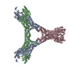
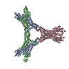
- PDB-6r7b: Crystal structure of Csx1 in complex with cyclic oligoadenylate c... -
+ Open data

- Basic information
| Entry | Database: PDB / ID: 6r7b | ||||||
|---|---|---|---|---|---|---|---|
| Title | Crystal structure of Csx1 in complex with cyclic oligoadenylate cOA4 conformation 1 | ||||||
 Components Components |
| ||||||
 Keywords Keywords | RNA BINDING PROTEIN / CRISPR ASSOCIATED PROTEIN CARF HEPN RNAse | ||||||
| Function / homology | SSO1389-like / : / CRISPR system endoribonuclease Csx1, CARF domain / Rossmann fold / 3-Layer(aba) Sandwich / Alpha Beta / RNA / CRISPR-associated (Cas) DxTHG family Function and homology information Function and homology information | ||||||
| Biological species |   Sulfolobus islandicus (archaea) Sulfolobus islandicus (archaea)synthetic construct (others) | ||||||
| Method |  X-RAY DIFFRACTION / SYNCHROTRON / MOLECULAR REPLACEMENT / Resolution: 3.12 Å X-RAY DIFFRACTION / SYNCHROTRON / MOLECULAR REPLACEMENT / Resolution: 3.12 Å | ||||||
 Authors Authors | Molina, R. / Montoya, G. / Sofos, N. / Stella, S. | ||||||
| Funding support |  Denmark, 1items Denmark, 1items
| ||||||
 Citation Citation | Journal: Nat Commun / Year: 2019 Title: Structure of Csx1-cOA complex reveals the basis of RNA decay in Type III-B CRISPR-Cas. Authors: Rafael Molina / Stefano Stella / Mingxia Feng / Nicholas Sofos / Vykintas Jauniskis / Irina Pozdnyakova / Blanca López-Méndez / Qunxin She / Guillermo Montoya /  Abstract: Type III CRISPR-Cas multisubunit complexes cleave ssRNA and ssDNA. These activities promote the generation of cyclic oligoadenylate (cOA), which activates associated CRISPR-Cas RNases from the ...Type III CRISPR-Cas multisubunit complexes cleave ssRNA and ssDNA. These activities promote the generation of cyclic oligoadenylate (cOA), which activates associated CRISPR-Cas RNases from the Csm/Csx families, triggering a massive RNA decay to provide immunity from genetic invaders. Here we present the structure of Sulfolobus islandicus (Sis) Csx1-cOA complex revealing the allosteric activation of its RNase activity. SisCsx1 is a hexamer built by a trimer of dimers. Each dimer forms a cOA binding site and a ssRNA catalytic pocket. cOA undergoes a conformational change upon binding in the second messenger binding site activating ssRNA degradation in the catalytic pockets. Activation is transmitted in an allosteric manner through an intermediate HTH domain, which joins the cOA and catalytic sites. The RNase functions in a sequential cooperative fashion, hydrolyzing phosphodiester bonds in 5'-C-C-3'. The degradation of cOA by Ring nucleases deactivates SisCsx1, suggesting that this enzyme could be employed in biotechnological applications. | ||||||
| History |
|
- Structure visualization
| Structure viewer | Molecule: MolmilJmol/JSmol |
|---|
- Downloads & links
-Download
| PDBx/mmCIF format | 6r7b.cif.gz | 559.6 KB | Display | PDBx/mmCIF format |
|---|---|---|---|---|
| PDB format | pdb6r7b.ent.gz | 470.4 KB | Display | PDB format |
| PDBx/mmJSON format | 6r7b.json.gz | Tree view | PDBx/mmJSON format | |
| Others |  Other downloads Other downloads |
-Validation report
| Arichive directory | https://data.pdbj.org/pub/pdb/validation_reports/r7/6r7bftp://data.pdbj.org/pub/pdb/validation_reports/r7/6r7b | HTTPS FTP |
|---|
-Related structure data
| Related structure data |  4691C 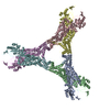 6qzqC  6qztSC  6r9rC C: citing same article ( S: Starting model for refinement |
|---|---|
| Similar structure data |

-Links
 PDBj
PDBj- Assembly
| Deposited unit | 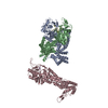
| ||||||||
|---|---|---|---|---|---|---|---|---|---|
| 1 | 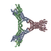
| ||||||||
| Unit cell |
|
-Components
| #1: Protein | Mass: 51859.395 Da / Num. of mol.: 3 Source method: isolated from a genetically manipulated source Source: (gene. exp.) Sulfolobus islandicus (archaea) / Gene: SiRe_0884 / Production host: Sulfolobus islandicus (archaea) / References: UniProt: F0NE21#2: RNA chain | Mass: 1271.866 Da / Num. of mol.: 2 / Source method: obtained synthetically Details: Although Chain D and E have the same composition, due to crystal symmetry chain E is just a half of D. Then chain D contains 4 adenosine monophosphate while chain E contains 2 adenosine monophosphates Source: (synth.) synthetic construct (others) #3: Water | ChemComp-HOH / |  Mass: 18.015 Da / Num. of mol.: 95 / Source method: isolated from a natural source / Formula: H2O Mass: 18.015 Da / Num. of mol.: 95 / Source method: isolated from a natural source / Formula: H2OHas protein modification | Y | |
|---|
-Experimental details
-Experiment
| Experiment | Method: X-RAY DIFFRACTION / Number of used crystals: 1 |
|---|
- Sample preparation
| Crystal | Density Matthews: 3.5 Å3/Da / Density % sol: 64.85 % / Description: PLATES |
|---|---|
| Crystal grow | Temperature: 293 K / Method: vapor diffusion, sitting drop / pH: 8 Details: 0.1M TRICINE PH=8.0, 0.35M NACL, 28% PEG1000, 10% GLYCEROL |
-Data collection
| Diffraction | Mean temperature: 100 K / Serial crystal experiment: N |
|---|---|
| Diffraction source | Source: SYNCHROTRON / Site: SLS  / Beamline: X06SA / Wavelength: 0.9785 Å / Beamline: X06SA / Wavelength: 0.9785 Å |
| Detector | Type: DECTRIS EIGER X 16M / Detector: PIXEL / Date: Nov 3, 2018 |
| Radiation | Protocol: SINGLE WAVELENGTH / Monochromatic (M) / Laue (L): M / Scattering type: x-ray |
| Radiation wavelength | Wavelength: 0.9785 Å / Relative weight: 1 |
| Reflection | Resolution: 3.12→178.63 Å / Num. obs: 24510 / % possible obs: 93 % / Redundancy: 11.6 % / Biso Wilson estimate: 115.32 Å2 / CC1/2: 0.99 / Net I/σ(I): 11.7 |
| Reflection shell | Resolution: 3.12→3.46 Å / Redundancy: 13.1 % / Mean I/σ(I) obs: 1.4 / Num. unique obs: 1226 / CC1/2: 0.57 |
- Processing
| Software |
| ||||||||||||||||||||||||||||||||||||||||||||||||||||||||||||||||||||||||||||||||||||||||||||||||||||||||||||||||||
|---|---|---|---|---|---|---|---|---|---|---|---|---|---|---|---|---|---|---|---|---|---|---|---|---|---|---|---|---|---|---|---|---|---|---|---|---|---|---|---|---|---|---|---|---|---|---|---|---|---|---|---|---|---|---|---|---|---|---|---|---|---|---|---|---|---|---|---|---|---|---|---|---|---|---|---|---|---|---|---|---|---|---|---|---|---|---|---|---|---|---|---|---|---|---|---|---|---|---|---|---|---|---|---|---|---|---|---|---|---|---|---|---|---|---|---|
| Refinement | Method to determine structure: MOLECULAR REPLACEMENT Starting model: 6QZT Resolution: 3.12→48.29 Å / Cor.coef. Fo:Fc: 0.948 / Cor.coef. Fo:Fc free: 0.923 / Cross valid method: THROUGHOUT / σ(F): 0 / SU Rfree Blow DPI: 0.556
| ||||||||||||||||||||||||||||||||||||||||||||||||||||||||||||||||||||||||||||||||||||||||||||||||||||||||||||||||||
| Displacement parameters | Biso mean: 131.32 Å2
| ||||||||||||||||||||||||||||||||||||||||||||||||||||||||||||||||||||||||||||||||||||||||||||||||||||||||||||||||||
| Refine analyze | Luzzati coordinate error obs: 0.41 Å | ||||||||||||||||||||||||||||||||||||||||||||||||||||||||||||||||||||||||||||||||||||||||||||||||||||||||||||||||||
| Refinement step | Cycle: LAST / Resolution: 3.12→48.29 Å
| ||||||||||||||||||||||||||||||||||||||||||||||||||||||||||||||||||||||||||||||||||||||||||||||||||||||||||||||||||
| Refine LS restraints |
| ||||||||||||||||||||||||||||||||||||||||||||||||||||||||||||||||||||||||||||||||||||||||||||||||||||||||||||||||||
| LS refinement shell | Resolution: 3.12→3.33 Å / Total num. of bins used: 50
| ||||||||||||||||||||||||||||||||||||||||||||||||||||||||||||||||||||||||||||||||||||||||||||||||||||||||||||||||||
| Refinement TLS params. | Method: refined / Refine-ID: X-RAY DIFFRACTION
| ||||||||||||||||||||||||||||||||||||||||||||||||||||||||||||||||||||||||||||||||||||||||||||||||||||||||||||||||||
| Refinement TLS group |
|
