 Movie
Movie Controller

[English] 日本語
 Yorodumi
- EMDB-4691: Reconstruction of the CRISPR-associated hexameric RNase Csx1 from... -
+ Open data

- Basic information
| Entry | Database: EMDB / ID: EMD-4691 | |||||||||
|---|---|---|---|---|---|---|---|---|---|---|
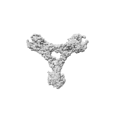
| Title | Reconstruction of the CRISPR-associated hexameric RNase Csx1 from Sulfolobus islandicus | |||||||||
 Map data Map data | Cryo-EM reconstruction of the Csx1 hexamer | |||||||||
 Sample Sample |
| |||||||||
| Function / homology | : / CRISPR system endoribonuclease Csx1, CARF domain / CRISPR-associated (Cas) DxTHG family Function and homology information Function and homology information | |||||||||
| Biological species |   Sulfolobus islandicus REY15A (archaea) Sulfolobus islandicus REY15A (archaea) | |||||||||
| Method | single particle reconstruction / cryo EM / Resolution: 3.62 Å | |||||||||
 Authors Authors | Montoya G / Sofos N / Molina R / Stella S | |||||||||
 Citation Citation | Journal: Nat Commun / Year: 2019 Title: Structure of Csx1-cOA complex reveals the basis of RNA decay in Type III-B CRISPR-Cas. Authors: Rafael Molina / Stefano Stella / Mingxia Feng / Nicholas Sofos / Vykintas Jauniskis / Irina Pozdnyakova / Blanca López-Méndez / Qunxin She / Guillermo Montoya /   Abstract: Type III CRISPR-Cas multisubunit complexes cleave ssRNA and ssDNA. These activities promote the generation of cyclic oligoadenylate (cOA), which activates associated CRISPR-Cas RNases from the ...Type III CRISPR-Cas multisubunit complexes cleave ssRNA and ssDNA. These activities promote the generation of cyclic oligoadenylate (cOA), which activates associated CRISPR-Cas RNases from the Csm/Csx families, triggering a massive RNA decay to provide immunity from genetic invaders. Here we present the structure of Sulfolobus islandicus (Sis) Csx1-cOA complex revealing the allosteric activation of its RNase activity. SisCsx1 is a hexamer built by a trimer of dimers. Each dimer forms a cOA binding site and a ssRNA catalytic pocket. cOA undergoes a conformational change upon binding in the second messenger binding site activating ssRNA degradation in the catalytic pockets. Activation is transmitted in an allosteric manner through an intermediate HTH domain, which joins the cOA and catalytic sites. The RNase functions in a sequential cooperative fashion, hydrolyzing phosphodiester bonds in 5'-C-C-3'. The degradation of cOA by Ring nucleases deactivates SisCsx1, suggesting that this enzyme could be employed in biotechnological applications. | |||||||||
| History |
|
- Structure visualization
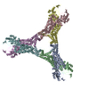
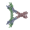
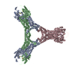
| Movie |
Movie viewer |
|---|---|
| Structure viewer | EM map: SurfViewMolmilJmol/JSmol |
| Supplemental images |
- Downloads & links
-EMDB archive
| Map data | emd_4691.map.gz | 226.3 MB | EMDB map data format | |
|---|---|---|---|---|
| Header (meta data) | emd-4691-v30.xmlemd-4691.xml | 11.5 KB 11.5 KB | Display Display | EMDB header |
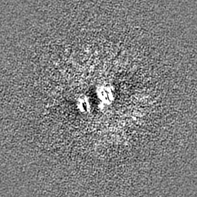
| Images |  emd_4691.png emd_4691.png | 58.3 KB | ||
| Archive directory |  http://ftp.pdbj.org/pub/emdb/structures/EMD-4691ftp://ftp.pdbj.org/pub/emdb/structures/EMD-4691 http://ftp.pdbj.org/pub/emdb/structures/EMD-4691ftp://ftp.pdbj.org/pub/emdb/structures/EMD-4691 | HTTPS FTP |
-Related structure data


-Links
| EMDB pages | EMDB (EBI/PDBe) / EMDataResource |
|---|
-Map
| File | Download / File: emd_4691.map.gz / Format: CCP4 / Size: 244.1 MB / Type: IMAGE STORED AS FLOATING POINT NUMBER (4 BYTES) | ||||||||||||||||||||||||||||||||||||||||||||||||||||||||||||
|---|---|---|---|---|---|---|---|---|---|---|---|---|---|---|---|---|---|---|---|---|---|---|---|---|---|---|---|---|---|---|---|---|---|---|---|---|---|---|---|---|---|---|---|---|---|---|---|---|---|---|---|---|---|---|---|---|---|---|---|---|---|
| Annotation | Cryo-EM reconstruction of the Csx1 hexamer | ||||||||||||||||||||||||||||||||||||||||||||||||||||||||||||
| Projections & slices | Image control
Images are generated by Spider. | ||||||||||||||||||||||||||||||||||||||||||||||||||||||||||||
| Voxel size | X=Y=Z: 1 Å | ||||||||||||||||||||||||||||||||||||||||||||||||||||||||||||
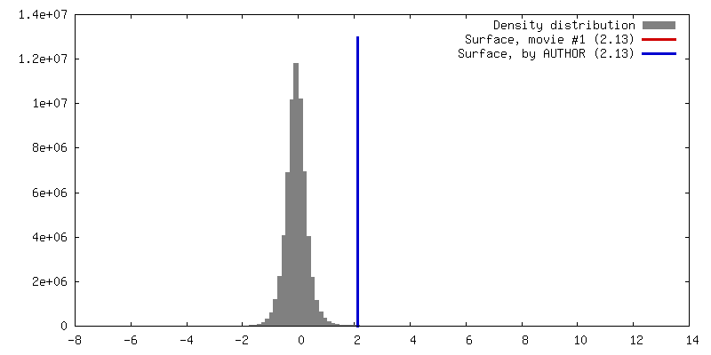
| Density |
| ||||||||||||||||||||||||||||||||||||||||||||||||||||||||||||
| Symmetry | Space group: 1 | ||||||||||||||||||||||||||||||||||||||||||||||||||||||||||||
| Details | EMDB XML:
CCP4 map header:
| ||||||||||||||||||||||||||||||||||||||||||||||||||||||||||||
 Z (Sec.)
 Y (Row.)
 X (Col.)


-Supplemental data
- Sample components
-Entire : Csx1 hexamer
| Entire | Name: Csx1 hexamer |
|---|---|
| Components |
|
-Supramolecule #1: Csx1 hexamer
| Supramolecule | Name: Csx1 hexamer / type: complex / ID: 1 / Parent: 0 |
|---|---|
| Source (natural) | Organism: Sulfolobus islandicus REY15A (archaea) / Location in cell: cytoplasm |
| Molecular weight | Experimental: 320 KDa |
-Experimental details
-Structure determination
| Method | cryo EM |
|---|---|
 Processing Processing | single particle reconstruction |
| Aggregation state | particle |
-Sample preparation
| Concentration | 0.5 mg/mL | |||||||||
|---|---|---|---|---|---|---|---|---|---|---|
| Buffer | pH: 8 Component:
| |||||||||
| Grid | Model: Quantifoil, UltrAuFoil / Material: GOLD / Mesh: 300 / Pretreatment - Type: GLOW DISCHARGE | |||||||||
| Vitrification | Cryogen name: ETHANE / Chamber humidity: 100 % / Chamber temperature: 277 K / Instrument: FEI VITROBOT MARK IV |
- Electron microscopy
| Microscope | FEI TITAN KRIOS |
|---|---|
| Image recording | Film or detector model: FEI FALCON III (4k x 4k) / Detector mode: COUNTING / Number grids imaged: 2 / Number real images: 2061 / Average electron dose: 50.0 e/Å2 Details: 808 exposures received 60 electrons pr square Angstrom. 1253 exposures received 40 electrons pr square Angstrom. |
| Electron beam | Acceleration voltage: 300 kV / Electron source:  FIELD EMISSION GUN FIELD EMISSION GUN |
| Electron optics | Illumination mode: FLOOD BEAM / Imaging mode: BRIGHT FIELD / Cs: 2.7 mm / Nominal defocus max: -3.0 µm / Nominal defocus min: -2.1 µm / Nominal magnification: 96000 |
| Sample stage | Specimen holder model: FEI TITAN KRIOS AUTOGRID HOLDER / Cooling holder cryogen: NITROGEN |
| Experimental equipment |  Model: Titan Krios / Image courtesy: FEI Company |
