 Movie
Movie Controller


+ Open data

- Basic information
| Entry | Database: PDB / ID: 6z6h | ||||||
|---|---|---|---|---|---|---|---|
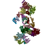
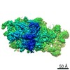
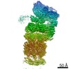
| Title | HDAC-DC | ||||||
 Components Components |
| ||||||
 Keywords Keywords | GENE REGULATION / Protein complex | ||||||
| Function / homology |  Function and homology information Function and homology informationHDA1 complex / HSF1 activation / HDACs deacetylate histones / histone deacetylase activity, hydrolytic mechanism / histone deacetylase / nucleosome array spacer activity / regulatory ncRNA-mediated gene silencing / SUMOylation of chromatin organization proteins / histone deacetylase complex / epigenetic regulation of gene expression ...HDA1 complex / HSF1 activation / HDACs deacetylate histones / histone deacetylase activity, hydrolytic mechanism / histone deacetylase / nucleosome array spacer activity / regulatory ncRNA-mediated gene silencing / SUMOylation of chromatin organization proteins / histone deacetylase complex / epigenetic regulation of gene expression / chromosome segregation / heterochromatin formation / chromatin remodeling / chromatin binding / regulation of transcription by RNA polymerase II / negative regulation of transcription by RNA polymerase II / positive regulation of transcription by RNA polymerase II / DNA binding / identical protein binding / nucleus / cytoplasm / cytosol Similarity search - Function | ||||||
| Biological species |  | ||||||
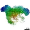
| Method | ELECTRON MICROSCOPY / single particle reconstruction / cryo EM / Resolution: 8.55 Å | ||||||
 Authors Authors | Lee, J.-H. / Bollschweiler, D. / Schaefer, T. / Huber, R. | ||||||
| Funding support |  Germany, 1items Germany, 1items
| ||||||
 Citation Citation | Journal: Sci Adv / Year: 2021 Title: Structural basis for the regulation of nucleosome recognition and HDAC activity by histone deacetylase assemblies. Authors: Jung-Hoon Lee / Daniel Bollschweiler / Tillman Schäfer / Robert Huber / Abstract: The chromatin-modifying histone deacetylases (HDACs) remove acetyl groups from acetyl-lysine residues in histone amino-terminal tails, thereby mediating transcriptional repression. Structural makeup ...The chromatin-modifying histone deacetylases (HDACs) remove acetyl groups from acetyl-lysine residues in histone amino-terminal tails, thereby mediating transcriptional repression. Structural makeup and mechanisms by which multisubunit HDAC complexes recognize nucleosomes remain elusive. Our cryo-electron microscopy structures of the yeast class II HDAC ensembles show that the HDAC protomer comprises a triangle-shaped assembly of stoichiometry Hda1-Hda2-Hda3, in which the active sites of the Hda1 dimer are freely accessible. We also observe a tetramer of protomers, where the nucleosome binding modules are inaccessible. Structural analysis of the nucleosome-bound complexes indicates how positioning of Hda1 adjacent to histone H2B affords HDAC catalysis. Moreover, it reveals how an intricate network of multiple contacts between a dimer of protomers and the nucleosome creates a platform for expansion of the HDAC activities. Our study provides comprehensive insight into the structural plasticity of the HDAC complex and its functional mechanism of chromatin modification. | ||||||
| History |
|
- Structure visualization
| Movie |
Movie viewer |
|---|---|
| Structure viewer | Molecule: MolmilJmol/JSmol |
- Downloads & links
-Download
| PDBx/mmCIF format | 6z6h.cif.gz | 861.9 KB | Display | PDBx/mmCIF format |
|---|---|---|---|---|
| PDB format | pdb6z6h.ent.gz | 687.6 KB | Display | PDB format |
| PDBx/mmJSON format | 6z6h.json.gz | Tree view | PDBx/mmJSON format | |
| Others |  Other downloads Other downloads |
-Validation report
| Arichive directory | https://data.pdbj.org/pub/pdb/validation_reports/z6/6z6hftp://data.pdbj.org/pub/pdb/validation_reports/z6/6z6h | HTTPS FTP |
|---|
-Related structure data
| Related structure data | 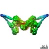 11094MC  6z6fC  6z6oC  6z6pC M: map data used to model this data C: citing same article ( |
|---|---|
| Similar structure data |


-Links
 PDBj
PDBj
- Assembly
| Deposited unit | 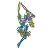
|
|---|---|
| 1 |
|
-Components
| #1: Protein | Mass: 74851.953 Da / Num. of mol.: 2 Source method: isolated from a genetically manipulated source Source: (gene. exp.) Gene: HDA1, YNL021W, N2819 / Production host:  #2: Protein | Mass: 76017.211 Da / Num. of mol.: 2 Source method: isolated from a genetically manipulated source Source: (gene. exp.) Gene: HDA1, YNL021W, N2819 / Production host: #3: Protein | Mass: 71915.297 Da / Num. of mol.: 2 Source method: isolated from a genetically manipulated source Source: (gene. exp.) Gene: HDA2, PLO2, YDR295C / Production host: #4: Protein | Mass: 63422.098 Da / Num. of mol.: 2 Source method: isolated from a genetically manipulated source Source: (gene. exp.) Gene: HDA3, PLO1, YPR179C / Production host: #5: Chemical | ChemComp-ZN /   Mass: 65.409 Da / Num. of mol.: 4 / Source method: obtained synthetically / Formula: Zn / Feature type: SUBJECT OF INVESTIGATION Mass: 65.409 Da / Num. of mol.: 4 / Source method: obtained synthetically / Formula: Zn / Feature type: SUBJECT OF INVESTIGATIONHas ligand of interest | Y | Has protein modification | Y | |
|---|
-Experimental details
-Experiment
| Experiment | Method: ELECTRON MICROSCOPY |
|---|---|
| EM experiment | Aggregation state: PARTICLE / 3D reconstruction method: single particle reconstruction |
- Sample preparation
| Component | Name: HDAC-DC / Type: COMPLEX / Entity ID: #1-#4 / Source: RECOMBINANT |
|---|---|
| Source (natural) | Organism: |
| Source (recombinant) | Organism: |
| Buffer solution | pH: 7.5 |
| Specimen | Embedding applied: NO / Shadowing applied: NO / Staining applied: NO / Vitrification applied: YES |
| Vitrification | Cryogen name: ETHANE |
- Electron microscopy imaging
| Microscopy | Model: TFS GLACIOS |
|---|---|
| Electron gun | Electron source:  FIELD EMISSION GUN / Accelerating voltage: 200 kV / Illumination mode: OTHER FIELD EMISSION GUN / Accelerating voltage: 200 kV / Illumination mode: OTHER |
| Electron lens | Mode: OTHER |
| Image recording | Electron dose: 62 e/Å2 / Film or detector model: GATAN K2 SUMMIT (4k x 4k) |
- Processing
| Software |
| ||||||||||||||||||||||||
|---|---|---|---|---|---|---|---|---|---|---|---|---|---|---|---|---|---|---|---|---|---|---|---|---|---|
| EM software |
| ||||||||||||||||||||||||
| CTF correction | Type: NONE | ||||||||||||||||||||||||
| Symmetry | Point symmetry: C2 (2 fold cyclic) | ||||||||||||||||||||||||
| 3D reconstruction | Resolution: 8.55 Å / Resolution method: FSC 0.143 CUT-OFF / Num. of particles: 11396 / Symmetry type: POINT | ||||||||||||||||||||||||
| Atomic model building | Details: Real space refinement | ||||||||||||||||||||||||
| Refinement | Cross valid method: NONE Stereochemistry target values: GeoStd + Monomer Library + CDL v1.2 | ||||||||||||||||||||||||
| Displacement parameters | Biso mean: 739.14 Å2 | ||||||||||||||||||||||||
| Refine LS restraints |
|
