[English] 日本語
Yorodumi
- EMDB-11797: HDAC-PC bound to nucleosome -

+
Open data


ID or keywords:

Loading...

-
Basic information

Entry
Database: EMDB / ID: EMD-11797
TitleHDAC-PC bound to nucleosome
Map dataHDAC-PC bound to nucleosome
Sample
  • Complex: HDAC-PC-Nuc
    • Complex: Histone deacetylase
    • Complex: Histone
    • Complex: DNA
Function / homology
Function and homology information


HDA1 complex / negative regulation of transcription by transcription factor localization / HSF1 activation / HDACs deacetylate histones / regulatory ncRNA-mediated gene silencing / histone deacetylase / SUMOylation of chromatin organization proteins / histone deacetylase activity / histone deacetylase complex / chromosome segregation ...HDA1 complex / negative regulation of transcription by transcription factor localization / HSF1 activation / HDACs deacetylate histones / regulatory ncRNA-mediated gene silencing / histone deacetylase / SUMOylation of chromatin organization proteins / histone deacetylase activity / histone deacetylase complex / chromosome segregation / structural constituent of chromatin / nucleosome / nucleosome assembly / cellular response to lipopolysaccharide / chromatin remodeling / protein heterodimerization activity / chromatin binding / regulation of transcription by RNA polymerase II / negative regulation of transcription by RNA polymerase II / positive regulation of transcription by RNA polymerase II / DNA binding / nucleoplasm / identical protein binding / nucleus / cytoplasm / cytosol
Similarity search - Function
Histone deacetylase class II, yeast / Arb2 domain / HDA1 complex subunit 2/3 / HDA1 complex subunit 3 / HDA1 complex subunit 2/3 superfamily / Arb2-like domain / Class II histone deacetylase complex subunits 2 and 3 / Histone deacetylase family / Histone deacetylase domain / Histone deacetylase domain superfamily ...Histone deacetylase class II, yeast / Arb2 domain / HDA1 complex subunit 2/3 / HDA1 complex subunit 3 / HDA1 complex subunit 2/3 superfamily / Arb2-like domain / Class II histone deacetylase complex subunits 2 and 3 / Histone deacetylase family / Histone deacetylase domain / Histone deacetylase domain superfamily / Histone deacetylase domain / Ureohydrolase domain superfamily / Histone H2B signature. / Histone H2B / Histone H2B / Histone H2A conserved site / Histone H2A signature. / Histone H2A, C-terminal domain / C-terminus of histone H2A / Histone H2A / Histone 2A / Histone H4, conserved site / Histone H4 signature. / Histone H4 / Histone H4 / TATA box binding protein associated factor / CENP-T/Histone H4, histone fold / Centromere kinetochore component CENP-T histone fold / TATA box binding protein associated factor (TAF), histone-like fold domain / Histone H3 signature 1. / Histone H3 signature 2. / Histone H3 / Histone H3/CENP-A / Histone H2A/H2B/H3 / Core histone H2A/H2B/H3/H4 / Histone-fold
Similarity search - Domain/homology
Histone H2B / Histone H2B / Histone H3 / Histone H2B 1.1 / Histone H2A type 1 / Histone deacetylase HDA1 / Histone H4 / Histone H3.2 / HDA1 complex subunit 3 / HDA1 complex subunit 2 / Histone H2A
Similarity search - Component
Biological speciesSaccharomyces cerevisiae (strain ATCC 204508 / S288c) (yeast) / Xenopus laevis (African clawed frog) / unidentified plasmid (others)
Methodsingle particle reconstruction / cryo EM / Resolution: 4.37 Å
AuthorsLee J-H / Bollschweiler D / Schaefer T / Huber R
Funding support Germany, 1 items
OrganizationGrant numberCountry
Max Planck Society Germany
Citation
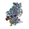
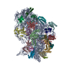
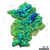
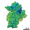
Journal: Sci Adv / Year: 2021
Title: Structural basis for the regulation of nucleosome recognition and HDAC activity by histone deacetylase assemblies.
Authors: Jung-Hoon Lee / Daniel Bollschweiler / Tillman Schäfer / Robert Huber /
Abstract: The chromatin-modifying histone deacetylases (HDACs) remove acetyl groups from acetyl-lysine residues in histone amino-terminal tails, thereby mediating transcriptional repression. Structural makeup ...The chromatin-modifying histone deacetylases (HDACs) remove acetyl groups from acetyl-lysine residues in histone amino-terminal tails, thereby mediating transcriptional repression. Structural makeup and mechanisms by which multisubunit HDAC complexes recognize nucleosomes remain elusive. Our cryo-electron microscopy structures of the yeast class II HDAC ensembles show that the HDAC protomer comprises a triangle-shaped assembly of stoichiometry Hda1-Hda2-Hda3, in which the active sites of the Hda1 dimer are freely accessible. We also observe a tetramer of protomers, where the nucleosome binding modules are inaccessible. Structural analysis of the nucleosome-bound complexes indicates how positioning of Hda1 adjacent to histone H2B affords HDAC catalysis. Moreover, it reveals how an intricate network of multiple contacts between a dimer of protomers and the nucleosome creates a platform for expansion of the HDAC activities. Our study provides comprehensive insight into the structural plasticity of the HDAC complex and its functional mechanism of chromatin modification.
History
DepositionSep 27, 2020-
Header (metadata) releaseMar 10, 2021-
Map releaseMar 10, 2021-
UpdateMar 10, 2021-
Current statusMar 10, 2021Processing site: PDBe / Status: Released

-
Structure visualization
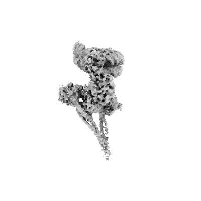
Movie
  • Surface view with section colored by density value
  • Surface level: 0.054
  • Imaged by UCSF Chimera
  • Download
  • Surface view colored by radius
  • Surface level: 0.054
  • Imaged by UCSF Chimera
  • Download
Movie viewer
Structure viewerEM map:
SurfViewMolmilJmol/JSmol
Supplemental images

Downloads & links

-
Map

FileDownload / File: emd_11797.map.gz / Format: CCP4 / Size: 600.7 MB / Type: IMAGE STORED AS FLOATING POINT NUMBER (4 BYTES)
AnnotationHDAC-PC bound to nucleosome
Voxel sizeX=Y=Z: 0.8512 Å
Density
Contour LevelBy AUTHOR: 0.054 / Movie #1: 0.054
Minimum - Maximum-0.10406329 - 0.28857774
Average (Standard dev.)-0.00015479307 (±0.009720516)
SymmetrySpace group: 1
Details

EMDB XML:

Map geometry
Axis orderXYZ
Origin000
Dimensions540540540
Spacing540540540
CellA=B=C: 459.64798 Å
α=β=γ: 90.0 °

CCP4 map header:

modeImage stored as Reals
Å/pix. X/Y/Z0.85120.85120.8512
M x/y/z540540540
origin x/y/z0.0000.0000.000
length x/y/z459.648459.648459.648
α/β/γ90.00090.00090.000
MAP C/R/S123
start NC/NR/NS000
NC/NR/NS540540540
D min/max/mean-0.1040.289-0.000

-
Supplemental data

-
Sample components

-
Entire : HDAC-PC-Nuc

EntireName: HDAC-PC-Nuc
Components
  • Complex: HDAC-PC-Nuc
    • Complex: Histone deacetylase
    • Complex: Histone
    • Complex: DNA

-
Supramolecule #1: HDAC-PC-Nuc

SupramoleculeName: HDAC-PC-Nuc / type: complex / ID: 1 / Parent: 0 / Macromolecule list: #1-#14

-
Supramolecule #2: Histone deacetylase

SupramoleculeName: Histone deacetylase / type: complex / ID: 2 / Parent: 1 / Macromolecule list: #1-#4
Source (natural)Organism: Saccharomyces cerevisiae (strain ATCC 204508 / S288c) (yeast)
Recombinant expressionOrganism: Escherichia coli (E. coli)

-
Supramolecule #3: Histone

SupramoleculeName: Histone / type: complex / ID: 3 / Parent: 1 / Macromolecule list: #5-#12
Source (natural)Organism: Xenopus laevis (African clawed frog)
Recombinant expressionOrganism: Escherichia coli (E. coli)

-
Supramolecule #4: DNA

SupramoleculeName: DNA / type: complex / ID: 4 / Parent: 1 / Macromolecule list: #13-#14
Source (natural)Organism: unidentified plasmid (others)
Recombinant expressionOrganism: synthetic construct (others)

-
Experimental details

-
Structure determination

Methodcryo EM
Processingsingle particle reconstruction
Aggregation stateparticle

-
Sample preparation

BufferpH: 7.5
VitrificationCryogen name: ETHANE

-
Electron microscopy

MicroscopeFEI TITAN KRIOS
Image recordingFilm or detector model: GATAN K3 BIOQUANTUM (6k x 4k) / Average electron dose: 77.2 e/Å2
Electron beamAcceleration voltage: 300 kV / Electron source: FIELD EMISSION GUN
Electron opticsIllumination mode: OTHER / Imaging mode: OTHER
Experimental equipment
Model: Titan Krios / Image courtesy: FEI Company

-
Image processing

Final reconstructionResolution.type: BY AUTHOR / Resolution: 4.37 Å / Resolution method: FSC 0.143 CUT-OFF / Software - Name: cisTEM (ver. 1) / Number images used: 41279
Initial angle assignmentType: OTHER
Final angle assignmentType: OTHER

-
Atomic model buiding 1

DetailsReal space refinement

+
About Yorodumi

-
News

-
Feb 9, 2022. New format data for meta-information of EMDB entries

New format data for meta-information of EMDB entries

  • Version 3 of the EMDB header file is now the official format.
  • The previous official version 1.9 will be removed from the archive.

Related info.:EMDB header

External links:wwPDB to switch to version 3 of the EMDB data model

-
Aug 12, 2020. Covid-19 info

Covid-19 info

URL: https://pdbjlvh1.pdbj.org/emnavi/covid19.php

New page: Covid-19 featured information page in EM Navigator.

Related info.:Covid-19 info / Mar 5, 2020. Novel coronavirus structure data

+
Mar 5, 2020. Novel coronavirus structure data

Novel coronavirus structure data

Related info.:Yorodumi Speices / Aug 12, 2020. Covid-19 info

External links:COVID-19 featured content - PDBj / Molecule of the Month (242):Coronavirus Proteases

+
Jan 31, 2019. EMDB accession codes are about to change! (news from PDBe EMDB page)

EMDB accession codes are about to change! (news from PDBe EMDB page)

  • The allocation of 4 digits for EMDB accession codes will soon come to an end. Whilst these codes will remain in use, new EMDB accession codes will include an additional digit and will expand incrementally as the available range of codes is exhausted. The current 4-digit format prefixed with “EMD-” (i.e. EMD-XXXX) will advance to a 5-digit format (i.e. EMD-XXXXX), and so on. It is currently estimated that the 4-digit codes will be depleted around Spring 2019, at which point the 5-digit format will come into force.
  • The EM Navigator/Yorodumi systems omit the EMD- prefix.

Related info.:Q: What is EMD? / ID/Accession-code notation in Yorodumi/EM Navigator

External links:EMDB Accession Codes are Changing Soon! / Contact to PDBj

+
Jul 12, 2017. Major update of PDB

Major update of PDB

  • wwPDB released updated PDB data conforming to the new PDBx/mmCIF dictionary.
  • This is a major update changing the version number from 4 to 5, and with Remediation, in which all the entries are updated.
  • In this update, many items about electron microscopy experimental information are reorganized (e.g. em_software).
  • Now, EM Navigator and Yorodumi are based on the updated data.

External links:wwPDB Remediation / Enriched Model Files Conforming to OneDep Data Standards Now Available in the PDB FTP Archive

-
Yorodumi

Thousand views of thousand structures

  • Yorodumi is a browser for structure data from EMDB, PDB, SASBDB, etc.
  • This page is also the successor to EM Navigator detail page, and also detail information page/front-end page for Omokage search.
  • The word "yorodu" (or yorozu) is an old Japanese word meaning "ten thousand". "mi" (miru) is to see.

Related info.:EMDB / PDB / SASBDB / Comparison of 3 databanks / Yorodumi Search / Aug 31, 2016. New EM Navigator & Yorodumi / Yorodumi Papers / Jmol/JSmol / Function and homology information / Changes in new EM Navigator and Yorodumi

Read more