[English] 日本語
Yorodumi
- PDB-6z1z: Structure of the anti-CD9 nanobody 4C8 -

+
Open data


ID or keywords:

Loading...

-
Basic information

Entry
Database: PDB / ID: 6z1z
TitleStructure of the anti-CD9 nanobody 4C8
ComponentsNanobody 4C8
KeywordsIMMUNE SYSTEM / nanobody / CD9-binding
Biological speciesLama glama (llama)
MethodX-RAY DIFFRACTION / SYNCHROTRON / MOLECULAR REPLACEMENT / Resolution: 1.7 Å
AuthorsNeviani, N. / Oosterheert, W. / Pearce, N.M. / Lutz, M. / Kroon-Batenburg, L.M.J. / Gros, P.
Funding support Netherlands, 2items
OrganizationGrant numberCountry
Netherlands Organisation for Scientific Research (NWO)731.015.201 Netherlands
Netherlands Organisation for Scientific Research (NWO)024.002.009 Netherlands
Citation
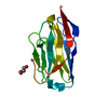
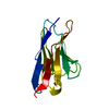
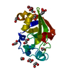
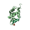
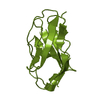
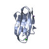
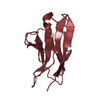
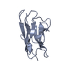
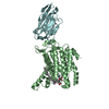
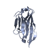
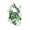
Journal: Life Sci Alliance / Year: 2020
Title: Implications for tetraspanin-enriched microdomain assembly based on structures of CD9 with EWI-F.
Authors: Wout Oosterheert / Katerina T Xenaki / Viviana Neviani / Wouter Pos / Sofia Doulkeridou / Jip Manshande / Nicholas M Pearce / Loes Mj Kroon-Batenburg / Martin Lutz / Paul Mp van Bergen En ...Authors: Wout Oosterheert / Katerina T Xenaki / Viviana Neviani / Wouter Pos / Sofia Doulkeridou / Jip Manshande / Nicholas M Pearce / Loes Mj Kroon-Batenburg / Martin Lutz / Paul Mp van Bergen En Henegouwen / Piet Gros /
Abstract: Tetraspanins are eukaryotic membrane proteins that contribute to a variety of signaling processes by organizing partner-receptor molecules in the plasma membrane. How tetraspanins bind and cluster ...Tetraspanins are eukaryotic membrane proteins that contribute to a variety of signaling processes by organizing partner-receptor molecules in the plasma membrane. How tetraspanins bind and cluster partner receptors into tetraspanin-enriched microdomains is unknown. Here, we present crystal structures of the large extracellular loop of CD9 bound to nanobodies 4C8 and 4E8 and, the cryo-EM structure of 4C8-bound CD9 in complex with its partner EWI-F. CD9-EWI-F displays a tetrameric arrangement with two central EWI-F molecules, dimerized through their ectodomains, and two CD9 molecules, one bound to each EWI-F transmembrane helix through CD9-helices h3 and h4. In the crystal structures, nanobodies 4C8 and 4E8 bind CD9 at loops C and D, which is in agreement with the 4C8 conformation in the CD9-EWI-F complex. The complex varies from nearly twofold symmetric (with the two CD9 copies nearly anti-parallel) to ca. 50° bent arrangements. This flexible arrangement of CD9-EWI-F with potential CD9 homo-dimerization at either end provides a "concatenation model" for forming short linear or circular assemblies, which may explain the occurrence of tetraspanin-enriched microdomains.
History
DepositionMay 14, 2020Deposition site: PDBE / Processing site: PDBE
Revision 1.0Sep 23, 2020Provider: repository / Type: Initial release
Revision 1.1Sep 30, 2020Group: Database references / Category: citation / citation_author
Item: _citation.country / _citation.journal_abbrev ..._citation.country / _citation.journal_abbrev / _citation.journal_id_CSD / _citation.journal_id_ISSN / _citation.journal_volume / _citation.pdbx_database_id_DOI / _citation.pdbx_database_id_PubMed / _citation.title / _citation.year
Revision 1.2Jan 24, 2024Group: Data collection / Database references / Refinement description
Category: chem_comp_atom / chem_comp_bond ...chem_comp_atom / chem_comp_bond / database_2 / pdbx_initial_refinement_model
Item: _database_2.pdbx_DOI / _database_2.pdbx_database_accession

-
Structure visualization

Structure viewerMolecule:
MolmilJmol/JSmol

Downloads & links

-
Assembly

Deposited unit
A: Nanobody 4C8
B: Nanobody 4C8


Theoretical massNumber of molelcules
Total (without water)28,1812
Polymers28,1812
Non-polymers00
Water2,522140
1
A: Nanobody 4C8


Theoretical massNumber of molelcules
Total (without water)14,0911
Polymers14,0911
Non-polymers00
Water181
TypeNameSymmetry operationNumber
identity operation1_555x,y,z1
2
B: Nanobody 4C8


Theoretical massNumber of molelcules
Total (without water)14,0911
Polymers14,0911
Non-polymers00
Water181
TypeNameSymmetry operationNumber
identity operation1_555x,y,z1
Unit cell
Length a, b, c (Å)48.901, 41.417, 56.853
Angle α, β, γ (deg.)90.000, 89.691, 90.000
Int Tables number4
Space group name H-MP1211
Space group name HallP2yb
Symmetry operation#1: x,y,z
#2: -x,y+1/2,-z

-
Components

#1: Antibody Nanobody 4C8


Mass: 14090.641 Da / Num. of mol.: 2
Source method: isolated from a genetically manipulated source
Source: (gene. exp.) Lama glama (llama) / Plasmid: pHEN6 / Production host: Escherichia coli BL21(DE3) (bacteria) / Variant (production host): Codon Plus (RIL)
#2: Water ChemComp-HOH / water


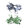
Mass: 18.015 Da / Num. of mol.: 140 / Source method: isolated from a natural source / Formula: H2O

-
Experimental details

-
Experiment

ExperimentMethod: X-RAY DIFFRACTION / Number of used crystals: 1

-
Sample preparation

CrystalDensity Matthews: 2.04 Å3/Da / Density % sol: 39.79 % / Description: Needle
Crystal growTemperature: 293 K / Method: vapor diffusion, sitting drop / pH: 7.5
Details: 0.1 M HEPES pH 7.5, 20% (w/v) PEG 8,000 The crystal was cryoprotected by soaking in reservoir solution supplemented with 30% (v/v) PEG 400
Temp details: Room temperature

-
Data collection

DiffractionMean temperature: 100 K / Ambient temp details: liquid nitrogen temperature / Serial crystal experiment: N
Diffraction sourceSource: SYNCHROTRON / Site: ESRF / Beamline: ID23-2 / Wavelength: 0.8731 Å
DetectorType: DECTRIS PILATUS3 X 2M / Detector: PIXEL / Date: Jun 16, 2018
RadiationProtocol: SINGLE WAVELENGTH / Monochromatic (M) / Laue (L): M / Scattering type: x-ray
Radiation wavelengthWavelength: 0.8731 Å / Relative weight: 1
ReflectionResolution: 1.7→24.64 Å / Num. obs: 23719 / % possible obs: 94.1 % / Redundancy: 3.4 % / CC1/2: 0.993 / Rmerge(I) obs: 0.129 / Rpim(I) all: 0.083 / Net I/σ(I): 4.1
Reflection shellResolution: 1.7→1.76 Å / Redundancy: 3.4 % / Rmerge(I) obs: 1.057 / Mean I/σ(I) obs: 0.8 / Num. unique obs: 2338 / CC1/2: 0.429 / Rpim(I) all: 0.677 / % possible all: 93.4

-
Processing

Software
NameVersionClassification
REFMAC5refinement
PHENIX1.14_3260refinement
EVAL15data reduction
EVAL15data scaling
PHASERphasing
RefinementMethod to determine structure: MOLECULAR REPLACEMENT
Starting model: 5IMK
Resolution: 1.7→24.63 Å / SU ML: 0.229 / Cross valid method: FREE R-VALUE / σ(F): 1.34 / Phase error: 25.3116 / Stereochemistry target values: CDL v1.2
RfactorNum. reflection% reflectionSelection details
Rfree0.231 1170 4.93 %random 5% of data
Rwork0.2011 22543 --
obs0.2027 23713 93.69 %-
Solvent computationShrinkage radii: 0.9 Å / VDW probe radii: 1.11 Å / Solvent model: FLAT BULK SOLVENT MODEL
Displacement parametersBiso mean: 25.27 Å2
Refinement stepCycle: LAST / Resolution: 1.7→24.63 Å
ProteinNucleic acidLigandSolventTotal
Num. atoms1772 0 0 140 1912
Refine LS restraints
Refine-IDTypeDev idealNumber
X-RAY DIFFRACTIONf_bond_d0.00231877
X-RAY DIFFRACTIONf_angle_d0.55072559
X-RAY DIFFRACTIONf_chiral_restr0.0434269
X-RAY DIFFRACTIONf_plane_restr0.0027331
X-RAY DIFFRACTIONf_dihedral_angle_d15.56361062
LS refinement shell
Resolution (Å)Rfactor RfreeNum. reflection RfreeRfactor RworkNum. reflection RworkRefine-ID% reflection obs (%)
1.7-1.780.30151450.32342787X-RAY DIFFRACTION93.29
1.78-1.870.30211390.26652797X-RAY DIFFRACTION93.71
1.87-1.990.2791460.23212801X-RAY DIFFRACTION93.85
1.99-2.140.24371580.21662827X-RAY DIFFRACTION94.37
2.14-2.360.20671230.19012787X-RAY DIFFRACTION93.03
2.36-2.70.2711440.19392848X-RAY DIFFRACTION94.5
2.7-3.40.22061500.18932858X-RAY DIFFRACTION94.77
3.4-24.630.19381650.17412838X-RAY DIFFRACTION92.12
Refinement TLS params.

Method: refined / Refine-ID: X-RAY DIFFRACTION

IDL112)L122)L132)L222)L232)L332)S11 (Å °)S12 (Å °)S13 (Å °)S21 (Å °)S22 (Å °)S23 (Å °)S31 (Å °)S32 (Å °)S33 (Å °)T112)T122)T132)T222)T232)T332)Origin x (Å)Origin y (Å)Origin z (Å)
11.59625212145-0.484816549043-0.0202135652141.23918430394-0.05874245238471.48191085490.04585927864980.116283992871-0.0739243924981-0.154270380471-0.10285072816-0.0346393398559-0.013341831777-0.0850469989348-0.02371642169030.118454616034-0.000340747667375-0.007613872722040.14284037776-0.01807361307380.13948354361812.0107785784-2.311163051250.964914243534
21.740592762840.290275487501-0.5462610666220.716921672831-0.05043087218652.352677421630.0523780362671-0.134554771420.1299636389860.0857386645128-0.07303334759560.069860574554-0.0191929562690.03926135985670.008429917456230.1348625969520.00396197610384-0.009031980052350.145336985536-0.01709281708040.14934820846812.111028108310.8400508743-26.7782902004
Refinement TLS group
IDRefine-IDRefine TLS-IDSelection details
1X-RAY DIFFRACTION1(chain A and resseq 1:121)
2X-RAY DIFFRACTION2(chain B and resseq 1:121)

+
About Yorodumi

-
News

-
Feb 9, 2022. New format data for meta-information of EMDB entries

New format data for meta-information of EMDB entries

  • Version 3 of the EMDB header file is now the official format.
  • The previous official version 1.9 will be removed from the archive.

Related info.:EMDB header

External links:wwPDB to switch to version 3 of the EMDB data model

-
Aug 12, 2020. Covid-19 info

Covid-19 info

URL: https://pdbjlvh1.pdbj.org/emnavi/covid19.php

New page: Covid-19 featured information page in EM Navigator.

Related info.:Covid-19 info / Mar 5, 2020. Novel coronavirus structure data

+
Mar 5, 2020. Novel coronavirus structure data

Novel coronavirus structure data

Related info.:Yorodumi Speices / Aug 12, 2020. Covid-19 info

External links:COVID-19 featured content - PDBj / Molecule of the Month (242):Coronavirus Proteases

+
Jan 31, 2019. EMDB accession codes are about to change! (news from PDBe EMDB page)

EMDB accession codes are about to change! (news from PDBe EMDB page)

  • The allocation of 4 digits for EMDB accession codes will soon come to an end. Whilst these codes will remain in use, new EMDB accession codes will include an additional digit and will expand incrementally as the available range of codes is exhausted. The current 4-digit format prefixed with “EMD-” (i.e. EMD-XXXX) will advance to a 5-digit format (i.e. EMD-XXXXX), and so on. It is currently estimated that the 4-digit codes will be depleted around Spring 2019, at which point the 5-digit format will come into force.
  • The EM Navigator/Yorodumi systems omit the EMD- prefix.

Related info.:Q: What is EMD? / ID/Accession-code notation in Yorodumi/EM Navigator

External links:EMDB Accession Codes are Changing Soon! / Contact to PDBj

+
Jul 12, 2017. Major update of PDB

Major update of PDB

  • wwPDB released updated PDB data conforming to the new PDBx/mmCIF dictionary.
  • This is a major update changing the version number from 4 to 5, and with Remediation, in which all the entries are updated.
  • In this update, many items about electron microscopy experimental information are reorganized (e.g. em_software).
  • Now, EM Navigator and Yorodumi are based on the updated data.

External links:wwPDB Remediation / Enriched Model Files Conforming to OneDep Data Standards Now Available in the PDB FTP Archive

-
Yorodumi

Thousand views of thousand structures

  • Yorodumi is a browser for structure data from EMDB, PDB, SASBDB, etc.
  • This page is also the successor to EM Navigator detail page, and also detail information page/front-end page for Omokage search.
  • The word "yorodu" (or yorozu) is an old Japanese word meaning "ten thousand". "mi" (miru) is to see.

Related info.:EMDB / PDB / SASBDB / Comparison of 3 databanks / Yorodumi Search / Aug 31, 2016. New EM Navigator & Yorodumi / Yorodumi Papers / Jmol/JSmol / Function and homology information / Changes in new EM Navigator and Yorodumi

Read more