 Movie
Movie Controller


+ Open data

- Basic information
| Entry | Database: PDB / ID: 6z20 | |||||||||
|---|---|---|---|---|---|---|---|---|---|---|
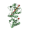
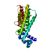
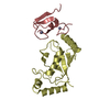
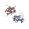
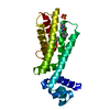
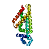
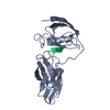
| Title | Structure of the EC2 domain of CD9 in complex with nanobody 4C8 | |||||||||
 Components Components |
| |||||||||
 Keywords Keywords | CELL ADHESION / Antibody-antigen complex / tetraspanin / CD9 / EC2 domain / nanobody | |||||||||
| Function / homology |  Function and homology information Function and homology informationAcrosome Reaction and Sperm:Oocyte Membrane Binding / myoblast fusion involved in skeletal muscle regeneration / sperm-egg recognition / fusion of sperm to egg plasma membrane involved in single fertilization / regulation of macrophage migration / paranodal junction assembly / glial cell migration / platelet alpha granule membrane / negative regulation of platelet aggregation / oligodendrocyte development ...Acrosome Reaction and Sperm:Oocyte Membrane Binding / myoblast fusion involved in skeletal muscle regeneration / sperm-egg recognition / fusion of sperm to egg plasma membrane involved in single fertilization / regulation of macrophage migration / paranodal junction assembly / glial cell migration / platelet alpha granule membrane / negative regulation of platelet aggregation / oligodendrocyte development / Uptake and function of diphtheria toxin / cellular response to low-density lipoprotein particle stimulus / clathrin-coated endocytic vesicle membrane / receptor internalization / platelet activation / integrin binding / endocytic vesicle membrane / Platelet degranulation / extracellular vesicle / cell population proliferation / cell adhesion / apical plasma membrane / negative regulation of cell population proliferation / external side of plasma membrane / focal adhesion / protein-containing complex / : / extracellular exosome / membrane / plasma membrane Similarity search - Function | |||||||||
| Biological species |  Homo sapiens (human) Homo sapiens (human) | |||||||||
| Method |  X-RAY DIFFRACTION / SYNCHROTRON / MOLECULAR REPLACEMENT / Resolution: 2.68 Å X-RAY DIFFRACTION / SYNCHROTRON / MOLECULAR REPLACEMENT / Resolution: 2.68 Å | |||||||||
 Authors Authors | Oosterheert, W. / Manshande, J. / Pearce, N.M. / Lutz, M. / Gros, P. | |||||||||
| Funding support |  Netherlands, 2items Netherlands, 2items
| |||||||||
 Citation Citation | Journal: Life Sci Alliance / Year: 2020 Title: Implications for tetraspanin-enriched microdomain assembly based on structures of CD9 with EWI-F. Authors: Wout Oosterheert / Katerina T Xenaki / Viviana Neviani / Wouter Pos / Sofia Doulkeridou / Jip Manshande / Nicholas M Pearce / Loes Mj Kroon-Batenburg / Martin Lutz / Paul Mp van Bergen En ...Authors: Wout Oosterheert / Katerina T Xenaki / Viviana Neviani / Wouter Pos / Sofia Doulkeridou / Jip Manshande / Nicholas M Pearce / Loes Mj Kroon-Batenburg / Martin Lutz / Paul Mp van Bergen En Henegouwen / Piet Gros / Abstract: Tetraspanins are eukaryotic membrane proteins that contribute to a variety of signaling processes by organizing partner-receptor molecules in the plasma membrane. How tetraspanins bind and cluster ...Tetraspanins are eukaryotic membrane proteins that contribute to a variety of signaling processes by organizing partner-receptor molecules in the plasma membrane. How tetraspanins bind and cluster partner receptors into tetraspanin-enriched microdomains is unknown. Here, we present crystal structures of the large extracellular loop of CD9 bound to nanobodies 4C8 and 4E8 and, the cryo-EM structure of 4C8-bound CD9 in complex with its partner EWI-F. CD9-EWI-F displays a tetrameric arrangement with two central EWI-F molecules, dimerized through their ectodomains, and two CD9 molecules, one bound to each EWI-F transmembrane helix through CD9-helices h3 and h4. In the crystal structures, nanobodies 4C8 and 4E8 bind CD9 at loops C and D, which is in agreement with the 4C8 conformation in the CD9-EWI-F complex. The complex varies from nearly twofold symmetric (with the two CD9 copies nearly anti-parallel) to ca. 50° bent arrangements. This flexible arrangement of CD9-EWI-F with potential CD9 homo-dimerization at either end provides a "concatenation model" for forming short linear or circular assemblies, which may explain the occurrence of tetraspanin-enriched microdomains. | |||||||||
| History |
|
- Structure visualization
| Structure viewer | Molecule: MolmilJmol/JSmol |
|---|
- Downloads & links
-Download
| PDBx/mmCIF format | 6z20.cif.gz | 283.5 KB | Display | PDBx/mmCIF format |
|---|---|---|---|---|
| PDB format | pdb6z20.ent.gz | 195.5 KB | Display | PDB format |
| PDBx/mmJSON format | 6z20.json.gz | Tree view | PDBx/mmJSON format | |
| Others |  Other downloads Other downloads |
-Validation report
| Arichive directory | https://data.pdbj.org/pub/pdb/validation_reports/z2/6z20ftp://data.pdbj.org/pub/pdb/validation_reports/z2/6z20 | HTTPS FTP |
|---|
-Related structure data
| Related structure data |  6rlrC  6z1vC 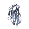 6z1zC  6rloS C: citing same article ( S: Starting model for refinement |
|---|---|
| Similar structure data |


-Links
 PDBj
PDBj


- Assembly
| Deposited unit | 
| ||||||||||||
|---|---|---|---|---|---|---|---|---|---|---|---|---|---|
| 1 | 
| ||||||||||||
| 2 | 
| ||||||||||||
| Unit cell |
|
-Components
| #1: Protein | Mass: 10129.433 Da / Num. of mol.: 2 Source method: isolated from a genetically manipulated source Source: (gene. exp.) Homo sapiens (human) / Gene: CD9, MIC3, TSPAN29, GIG2 / Plasmid: 107.03 / Cell (production host): EMBRYONIC / Cell line (production host): HEK293EBNA / Organ (production host): KIDNEY / Production host: Homo sapiens (human) / References: UniProt: P21926#2: Antibody | Mass: 14090.641 Da / Num. of mol.: 2 Source method: isolated from a genetically manipulated source Source: (gene. exp.)  #3: Chemical |   Mass: 92.094 Da / Num. of mol.: 2 / Source method: obtained synthetically / Formula: C3H8O3 Mass: 92.094 Da / Num. of mol.: 2 / Source method: obtained synthetically / Formula: C3H8O3#4: Chemical | ChemComp-CL / |   Mass: 35.453 Da / Num. of mol.: 1 / Source method: obtained synthetically / Formula: Cl Mass: 35.453 Da / Num. of mol.: 1 / Source method: obtained synthetically / Formula: Cl#5: Water | ChemComp-HOH / |  Mass: 18.015 Da / Num. of mol.: 6 / Source method: isolated from a natural source / Formula: H2O Mass: 18.015 Da / Num. of mol.: 6 / Source method: isolated from a natural source / Formula: H2OHas ligand of interest | N | Has protein modification | Y | |
|---|
-Experimental details
-Experiment
| Experiment | Method: X-RAY DIFFRACTION / Number of used crystals: 1 |
|---|
- Sample preparation
| Crystal | Density Matthews: 3.06 Å3/Da / Density % sol: 59.85 % / Description: needle |
|---|---|
| Crystal grow | Temperature: 293 K / Method: vapor diffusion, sitting drop / pH: 5.6 Details: 0.095 M sodium citrate pH 5.6, 5% (v/v) glycerol, 19% (v/v) isopropanol, 20% (w/v) PEG 4,000. The crystal was cryoprotected by soaking in reservoir solution supplemented with 25% glycerol (final concentration). Temp details: room temperature |
-Data collection
| Diffraction | Mean temperature: 100 K / Ambient temp details: liquid nitrogen temperature / Serial crystal experiment: N |
|---|---|
| Diffraction source | Source: SYNCHROTRON / Site: Diamond  / Beamline: I03 / Wavelength: 0.9763 Å / Beamline: I03 / Wavelength: 0.9763 Å |
| Detector | Type: DECTRIS EIGER2 XE 16M / Detector: PIXEL / Date: May 17, 2019 |
| Radiation | Protocol: SINGLE WAVELENGTH / Monochromatic (M) / Laue (L): M / Scattering type: x-ray |
| Radiation wavelength | Wavelength: 0.9763 Å / Relative weight: 1 |
| Reflection | Resolution: 2.68→88.49 Å / Num. obs: 14256 / % possible obs: 93.9 % / Redundancy: 6.7 % / Biso Wilson estimate: 66.57 Å2 / CC1/2: 0.997 / Rmerge(I) obs: 0.13 / Rpim(I) all: 0.05 / Net I/σ(I): 9.5 |
| Reflection shell | Resolution: 2.68→2.83 Å / Redundancy: 7.3 % / Rmerge(I) obs: 1.78 / Mean I/σ(I) obs: 1.1 / Num. unique obs: 750 / CC1/2: 0.507 / Rpim(I) all: 0.71 / % possible all: 55.8 |
- Processing
| Software |
| ||||||||||||||||||||||||||||||||||||||||||
|---|---|---|---|---|---|---|---|---|---|---|---|---|---|---|---|---|---|---|---|---|---|---|---|---|---|---|---|---|---|---|---|---|---|---|---|---|---|---|---|---|---|---|---|
| Refinement | Method to determine structure: MOLECULAR REPLACEMENT Starting model: 6RLO Resolution: 2.68→60.71 Å / SU ML: 0.3054 / Cross valid method: FREE R-VALUE / σ(F): 1.34 / Phase error: 29.879 / Stereochemistry target values: CDL v1.2
| ||||||||||||||||||||||||||||||||||||||||||
| Solvent computation | Shrinkage radii: 0.9 Å / VDW probe radii: 1.11 Å / Solvent model: FLAT BULK SOLVENT MODEL | ||||||||||||||||||||||||||||||||||||||||||
| Displacement parameters | Biso mean: 68.91 Å2 | ||||||||||||||||||||||||||||||||||||||||||
| Refinement step | Cycle: LAST / Resolution: 2.68→60.71 Å
| ||||||||||||||||||||||||||||||||||||||||||
| Refine LS restraints |
| ||||||||||||||||||||||||||||||||||||||||||
| LS refinement shell |
| ||||||||||||||||||||||||||||||||||||||||||
| Refinement TLS params. | Method: refined / Origin x: 20.6220696969 Å / Origin y: 11.8145232372 Å / Origin z: -22.5004079045 Å
| ||||||||||||||||||||||||||||||||||||||||||
| Refinement TLS group | Selection details: all |
