Netherlands Organisation for Scientific Research (NWO)
731.015.201
オランダ
Netherlands Organisation for Scientific Research (NWO)
024.002.009
オランダ
引用
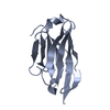
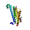
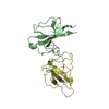
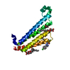
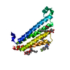
ジャーナル: Life Sci Alliance / 年: 2020 タイトル: Implications for tetraspanin-enriched microdomain assembly based on structures of CD9 with EWI-F. 著者: Wout Oosterheert / Katerina T Xenaki / Viviana Neviani / Wouter Pos / Sofia Doulkeridou / Jip Manshande / Nicholas M Pearce / Loes Mj Kroon-Batenburg / Martin Lutz / Paul Mp van Bergen En ...著者: Wout Oosterheert / Katerina T Xenaki / Viviana Neviani / Wouter Pos / Sofia Doulkeridou / Jip Manshande / Nicholas M Pearce / Loes Mj Kroon-Batenburg / Martin Lutz / Paul Mp van Bergen En Henegouwen / Piet Gros / 要旨: Tetraspanins are eukaryotic membrane proteins that contribute to a variety of signaling processes by organizing partner-receptor molecules in the plasma membrane. How tetraspanins bind and cluster ...Tetraspanins are eukaryotic membrane proteins that contribute to a variety of signaling processes by organizing partner-receptor molecules in the plasma membrane. How tetraspanins bind and cluster partner receptors into tetraspanin-enriched microdomains is unknown. Here, we present crystal structures of the large extracellular loop of CD9 bound to nanobodies 4C8 and 4E8 and, the cryo-EM structure of 4C8-bound CD9 in complex with its partner EWI-F. CD9-EWI-F displays a tetrameric arrangement with two central EWI-F molecules, dimerized through their ectodomains, and two CD9 molecules, one bound to each EWI-F transmembrane helix through CD9-helices h3 and h4. In the crystal structures, nanobodies 4C8 and 4E8 bind CD9 at loops C and D, which is in agreement with the 4C8 conformation in the CD9-EWI-F complex. The complex varies from nearly twofold symmetric (with the two CD9 copies nearly anti-parallel) to ca. 50° bent arrangements. This flexible arrangement of CD9-EWI-F with potential CD9 homo-dimerization at either end provides a "concatenation model" for forming short linear or circular assemblies, which may explain the occurrence of tetraspanin-enriched microdomains.
構造決定の手法: 分子置換 開始モデル: Structure of CD9EC2 bound to nanobody 4C8 解像度: 1.33→44.72 Å / SU ML: 0.1369 / 交差検証法: FREE R-VALUE / σ(F): 1.35 / 位相誤差: 25.1549 / 立体化学のターゲット値: CDL v1.2 詳細: For the CD9EC2 - 4E8 dataset, the autoprocessed and anisotropical-truncated (autoproc-staraniso) reflection data file provided by DLS was employed. The structure was solved by molecular ...詳細: For the CD9EC2 - 4E8 dataset, the autoprocessed and anisotropical-truncated (autoproc-staraniso) reflection data file provided by DLS was employed. The structure was solved by molecular replacement using PHASER with the CD9EC2 - 4C8 structure as search model. The 4C8 residues were replaced with the corresponding 4E8 residues and the CDR regions of the nanobody were manually built in Coot. The structure was then iteratively refined using Refmac5 or Phenix, alternated with model improvement in COOT. The final refinement in Phenix yielded Rwork/Rfree = 15.1/19.0%
Rfactor
反射数
%反射
Rfree
0.1898
1570
4.9 %
Rwork
0.1512
30472
-
obs
0.1531
32042
69.74 %
溶媒の処理
減衰半径: 0.9 Å / VDWプローブ半径: 1.11 Å / 溶媒モデル: FLAT BULK SOLVENT MODEL
 ムービー
ムービー コントローラー


 データを開く
 基本情報
基本情報 要素
要素 キーワード
キーワード 機能・相同性情報
 Homo sapiens (ヒト)
 X線回折 /
  データ登録者
 オランダ, 2件
  引用
引用 構造の表示
構造の表示 ダウンロードとリンク
 その他のダウンロード


 PDBj
PDBj


 集合体
集合体


 分子量: 150.173 Da / 分子数: 1 / 由来タイプ: 合成 / 式: C6H14O4
 分子量: 62.068 Da / 分子数: 1 / 由来タイプ: 合成 / 式: C2H6O2
 分子量: 60.052 Da / 分子数: 1 / 由来タイプ: 合成 / 式: C2H4O2
 試料調製
試料調製 / ビームライン: I04-1 / 波長: 0.9159 Å
 解析
解析