| Entry | Database: PDB / ID: 4yl1
|
|---|
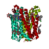
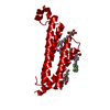
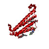
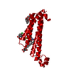
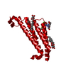
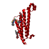
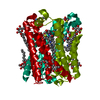
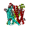
| Title | Crystal Structures of mPGES-1 Inhibitor Complexes |
|---|
 Components Components | Prostaglandin E synthase |
|---|
 Keywords Keywords | ISOMERASE/ISOMERASE INHIBITOR / Inhibitor / Inflammation / Prostaglandin / ISOMERASE-ISOMERASE INHIBITOR complex |
|---|
| Function / homology |  Function and homology information Function and homology information
prostaglandin-E synthase / prostaglandin-E synthase activity / regulation of fever generation / prostaglandin-D synthase activity / glutathione binding / cyclooxygenase pathway / Synthesis of Prostaglandins (PG) and Thromboxanes (TX) / glutathione peroxidase activity / prostaglandin biosynthetic process / prostaglandin metabolic process ...prostaglandin-E synthase / prostaglandin-E synthase activity / regulation of fever generation / prostaglandin-D synthase activity / glutathione binding / cyclooxygenase pathway / Synthesis of Prostaglandins (PG) and Thromboxanes (TX) / glutathione peroxidase activity / prostaglandin biosynthetic process / prostaglandin metabolic process / nuclear envelope lumen / glutathione transferase / glutathione transferase activity / Oxidoreductases; Acting on a peroxide as acceptor; Peroxidases / sensory perception of pain / regulation of inflammatory response / endoplasmic reticulum membrane / perinuclear region of cytoplasm / signal transduction / endoplasmic reticulum / membraneSimilarity search - Function Microsomal glutathione S-transferase 1-like / Membrane associated eicosanoid/glutathione metabolism-like domain / Membrane-associated, eicosanoid/glutathione metabolism (MAPEG) protein / Membrane associated eicosanoid/glutathione metabolism-like domain superfamily / MAPEG family / Four Helix Bundle (Hemerythrin (Met), subunit A) / Up-down Bundle / Mainly AlphaSimilarity search - Domain/homology |
|---|
| Biological species |  Homo sapiens (human) Homo sapiens (human) |
|---|
| Method |  X-RAY DIFFRACTION / SYNCHROTRON / MOLECULAR REPLACEMENT / molecular replacement / Resolution: 1.41 Å X-RAY DIFFRACTION / SYNCHROTRON / MOLECULAR REPLACEMENT / molecular replacement / Resolution: 1.41 Å |
|---|
 Authors Authors | Luz, J.G. / Antonysamy, S. / Kuklish, S.L. / Fisher, M.J. |
|---|
 Citation Citation | Journal: J.Med.Chem. / Year: 2015
Title: Crystal Structures of mPGES-1 Inhibitor Complexes Form a Basis for the Rational Design of Potent Analgesic and Anti-Inflammatory Therapeutics.
Authors: Luz, J.G. / Antonysamy, S. / Kuklish, S.L. / Condon, B. / Lee, M.R. / Allison, D. / Yu, X.P. / Chandrasekhar, S. / Backer, R. / Zhang, A. / Russell, M. / Chang, S.S. / Harvey, A. / Sloan, A.V. / Fisher, M.J. |
|---|
| History | | Deposition | Mar 4, 2015 | Deposition site: RCSB / Processing site: RCSB |
|---|
| Revision 1.0 | Jun 10, 2015 | Provider: repository / Type: Initial release |
|---|
| Revision 1.1 | Jul 8, 2015 | Group: Database references |
|---|
| Revision 1.2 | Nov 22, 2017 | Group: Database references / Derived calculations ...Database references / Derived calculations / Refinement description / Source and taxonomy / Structure summary
Category: citation / entity_src_gen ...citation / entity_src_gen / pdbx_struct_assembly / pdbx_struct_assembly_prop / pdbx_struct_oper_list / software / struct_keywords
Item: _citation.journal_id_CSD / _entity_src_gen.pdbx_alt_source_flag ..._citation.journal_id_CSD / _entity_src_gen.pdbx_alt_source_flag / _pdbx_struct_assembly.oligomeric_details / _pdbx_struct_assembly_prop.type / _pdbx_struct_assembly_prop.value / _pdbx_struct_oper_list.symmetry_operation / _software.classification / _struct_keywords.text |
|---|
| Revision 1.3 | Jul 29, 2020 | Group: Data collection / Derived calculations / Structure summary
Category: chem_comp / entity ...chem_comp / entity / pdbx_chem_comp_identifier / pdbx_entity_nonpoly / struct_site / struct_site_gen
Item: _chem_comp.mon_nstd_flag / _chem_comp.name ..._chem_comp.mon_nstd_flag / _chem_comp.name / _chem_comp.type / _entity.pdbx_description / _pdbx_entity_nonpoly.name
Description: Carbohydrate remediation / Provider: repository / Type: Remediation |
|---|
| Revision 1.4 | Feb 28, 2024 | Group: Data collection / Database references / Structure summary
Category: chem_comp / chem_comp_atom ...chem_comp / chem_comp_atom / chem_comp_bond / database_2
Item: _chem_comp.pdbx_synonyms / _database_2.pdbx_DOI / _database_2.pdbx_database_accession |
|---|
|
|---|
 Movie
Movie Controller


 Open data
 Basic information
 Structure visualization
 Downloads & links
 Other downloads


 PDBj
PDBj


 Assembly


 Spodoptera frugiperda (fall armyworm) / References: UniProt: O14684, prostaglandin-E synthase
 Type: D-saccharide / Mass: 292.369 Da / Num. of mol.: 2


 Mass: 427.535 Da / Num. of mol.: 1 / Source method: obtained synthetically / Formula: C28H29NO3
 Mass: 106.120 Da / Num. of mol.: 1 / Source method: obtained synthetically / Formula: C4H10O3
 Mass: 307.323 Da / Num. of mol.: 1 / Source method: obtained synthetically / Formula: C10H17N3O6S
 Mass: 194.226 Da / Num. of mol.: 1 / Source method: obtained synthetically / Formula: C8H18O5 / Comment: precipitant*YM
 Sample preparation
 / Beamline: 31-ID / Wavelength: 0.91986 Å
 Processing