 Movie
Movie Controller

[English] 日本語
 Yorodumi
- PDB-6kl5: Structure of The N-terminal domain of Middle East respiratory syn... -
+ Open data

- Basic information
| Entry | Database: PDB / ID: 6kl5 | ||||||
|---|---|---|---|---|---|---|---|
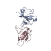
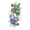
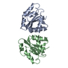
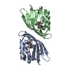
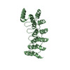
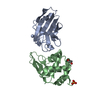
| Title | Structure of The N-terminal domain of Middle East respiratory syndrome coronavirus Nucleocapsid Protein complexed with Benzyl 2-(Hydroxymethyl)-1-Indolinecarboxylate | ||||||
 Components Components | Nucleoprotein | ||||||
 Keywords Keywords | VIRAL PROTEIN / Middle East respiratory syndrome coronavirus / nucleocapsid protein / N-terminal domain / Benzyl 2-(Hydroxymethyl)-1-Indoline carboxylate / VIRUS | ||||||
| Function / homology |  Function and homology information Function and homology informationviral RNA genome packaging / negative regulation of interferon-beta production / viral capsid / host cell / viral nucleocapsid / host cell endoplasmic reticulum-Golgi intermediate compartment / host cell Golgi apparatus / ribonucleoprotein complex / RNA binding Similarity search - Function | ||||||
| Biological species |  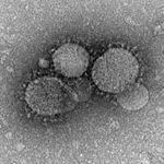 Middle East respiratory syndrome-related coronavirus Middle East respiratory syndrome-related coronavirus | ||||||
| Method |  X-RAY DIFFRACTION / SYNCHROTRON / MOLECULAR REPLACEMENT / Resolution: 3.09 Å X-RAY DIFFRACTION / SYNCHROTRON / MOLECULAR REPLACEMENT / Resolution: 3.09 Å | ||||||
 Authors Authors | Hou, M.H. / Lin, S.M. / Hsu, J.N. / Wang, Y.S. | ||||||
 Citation Citation | Journal: J.Med.Chem. / Year: 2020 Title: Structure-Based Stabilization of Non-native Protein-Protein Interactions of Coronavirus Nucleocapsid Proteins in Antiviral Drug Design. Authors: Lin, S.M. / Lin, S.C. / Hsu, J.N. / Chang, C.K. / Chien, C.M. / Wang, Y.S. / Wu, H.Y. / Jeng, U.S. / Kehn-Hall, K. / Hou, M.H. | ||||||
| History |
|
- Structure visualization
| Structure viewer | Molecule: MolmilJmol/JSmol |
|---|
- Downloads & links
-Download
| PDBx/mmCIF format | 6kl5.cif.gz | 100.7 KB | Display | PDBx/mmCIF format |
|---|---|---|---|---|
| PDB format | pdb6kl5.ent.gz | 75.4 KB | Display | PDB format |
| PDBx/mmJSON format | 6kl5.json.gz | Tree view | PDBx/mmJSON format | |
| Others |  Other downloads Other downloads |
-Validation report
| Arichive directory | https://data.pdbj.org/pub/pdb/validation_reports/kl/6kl5ftp://data.pdbj.org/pub/pdb/validation_reports/kl/6kl5 | HTTPS FTP |
|---|
-Related structure data
| Related structure data |  6kl2C  6kl6C  4j3kS S: Starting model for refinement C: citing same article ( |
|---|---|
| Similar structure data |


-Links
 PDBj
PDBj- Assembly
| Deposited unit | 
| ||||||||
|---|---|---|---|---|---|---|---|---|---|
| 1 | 
| ||||||||
| 2 | 
| ||||||||
| Unit cell |
|
-Components
| #1: Protein | Mass: 15855.566 Da / Num. of mol.: 4 / Fragment: N-terminal domain Source method: isolated from a genetically manipulated source Source: (gene. exp.) Middle East respiratory syndrome-related coronavirusProduction host:  #2: Chemical | ChemComp-DJO / ( |   Mass: 283.322 Da / Num. of mol.: 1 / Source method: obtained synthetically / Formula: C17H17NO3 / Feature type: SUBJECT OF INVESTIGATION Mass: 283.322 Da / Num. of mol.: 1 / Source method: obtained synthetically / Formula: C17H17NO3 / Feature type: SUBJECT OF INVESTIGATIONHas ligand of interest | Y | |
|---|
-Experimental details
-Experiment
| Experiment | Method: X-RAY DIFFRACTION / Number of used crystals: 1 |
|---|
- Sample preparation
| Crystal |
| |||||||||
|---|---|---|---|---|---|---|---|---|---|---|
| Crystal grow | Temperature: 293 K / Method: vapor diffusion, sitting drop Details: 5 mg/mL protein, 25 mM Tris-HCl, pH 7.5, 75 mM NaCl, 70 mM MES, pH5.5, 37.5 M (NH4)2SO4, 14.5 % PEG 3350, 1 mM NaBr, 1mM |
-Data collection
| Diffraction | Mean temperature: 110 K / Serial crystal experiment: N | |||||||||||||||||||||||||||||||||||||||||||||||||||||||||||||||||||||||||||||||||||||||||||||||||||
|---|---|---|---|---|---|---|---|---|---|---|---|---|---|---|---|---|---|---|---|---|---|---|---|---|---|---|---|---|---|---|---|---|---|---|---|---|---|---|---|---|---|---|---|---|---|---|---|---|---|---|---|---|---|---|---|---|---|---|---|---|---|---|---|---|---|---|---|---|---|---|---|---|---|---|---|---|---|---|---|---|---|---|---|---|---|---|---|---|---|---|---|---|---|---|---|---|---|---|---|---|
| Diffraction source | Source: SYNCHROTRON / Site: NSRRC  / Beamline: BL13C1 / Wavelength: 0.97622 Å / Beamline: BL13C1 / Wavelength: 0.97622 Å | |||||||||||||||||||||||||||||||||||||||||||||||||||||||||||||||||||||||||||||||||||||||||||||||||||
| Detector | Type: RAYONIX MX300HE / Detector: CCD / Date: Sep 26, 2015 | |||||||||||||||||||||||||||||||||||||||||||||||||||||||||||||||||||||||||||||||||||||||||||||||||||
| Radiation | Protocol: SINGLE WAVELENGTH / Monochromatic (M) / Laue (L): M / Scattering type: x-ray | |||||||||||||||||||||||||||||||||||||||||||||||||||||||||||||||||||||||||||||||||||||||||||||||||||
| Radiation wavelength | Wavelength: 0.97622 Å / Relative weight: 1 | |||||||||||||||||||||||||||||||||||||||||||||||||||||||||||||||||||||||||||||||||||||||||||||||||||
| Reflection | Resolution: 3.076→30 Å / Num. obs: 12334 / % possible obs: 96.1 % / Redundancy: 3.5 % / Rmerge(I) obs: 0.137 / Rpim(I) all: 0.084 / Rrim(I) all: 0.161 / Χ2: 0.928 / Net I/σ(I): 5.6 | |||||||||||||||||||||||||||||||||||||||||||||||||||||||||||||||||||||||||||||||||||||||||||||||||||
| Reflection shell | Diffraction-ID: 1
|
- Processing
| Software |
| |||||||||||||||||||||||||||||||||||||||||||||
|---|---|---|---|---|---|---|---|---|---|---|---|---|---|---|---|---|---|---|---|---|---|---|---|---|---|---|---|---|---|---|---|---|---|---|---|---|---|---|---|---|---|---|---|---|---|---|
| Refinement | Method to determine structure: MOLECULAR REPLACEMENT Starting model: 4J3K Resolution: 3.09→26.289 Å / SU ML: 0.48 / Cross valid method: THROUGHOUT / σ(F): 1.45 / Phase error: 30.29
| |||||||||||||||||||||||||||||||||||||||||||||
| Solvent computation | Shrinkage radii: 0.9 Å / VDW probe radii: 1.11 Å | |||||||||||||||||||||||||||||||||||||||||||||
| Displacement parameters | Biso max: 99.65 Å2 / Biso mean: 54.0869 Å2 / Biso min: 24.13 Å2 | |||||||||||||||||||||||||||||||||||||||||||||
| Refinement step | Cycle: final / Resolution: 3.09→26.289 Å
| |||||||||||||||||||||||||||||||||||||||||||||
| LS refinement shell | Refine-ID: X-RAY DIFFRACTION / Rfactor Rfree error: 0 / Rfactor Rwork: 0.2594
|
