 Movie
Movie Controller

[English] 日本語
 Yorodumi
- PDB-2itj: Origin binding domain of the SV40 large T antigen (residues 131-2... -
+ Open data

- Basic information
| Entry | Database: PDB / ID: 2itj | ||||||
|---|---|---|---|---|---|---|---|
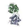
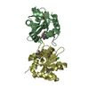
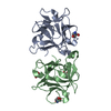
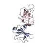
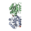
| Title | Origin binding domain of the SV40 large T antigen (residues 131-259). P212121 crystal form | ||||||
 Components Components | large T antigen | ||||||
 Keywords Keywords | DNA BINDING PROTEIN | ||||||
| Function / homology |  Function and homology information Function and homology informationsymbiont-mediated suppression of host JAK-STAT cascade via inhibition of JAK1 activity / bidirectional double-stranded viral DNA replication / viral DNA genome replication / symbiont-mediated perturbation of host cell cycle G1/S transition checkpoint / 3'-5' DNA helicase activity / DNA 3'-5' helicase / DNA replication origin binding / single-stranded DNA binding / double-stranded DNA binding / symbiont-mediated perturbation of host ubiquitin-like protein modification ...symbiont-mediated suppression of host JAK-STAT cascade via inhibition of JAK1 activity / bidirectional double-stranded viral DNA replication / viral DNA genome replication / symbiont-mediated perturbation of host cell cycle G1/S transition checkpoint / 3'-5' DNA helicase activity / DNA 3'-5' helicase / DNA replication origin binding / single-stranded DNA binding / double-stranded DNA binding / symbiont-mediated perturbation of host ubiquitin-like protein modification / DNA replication / symbiont-mediated suppression of host innate immune response / symbiont-mediated suppression of host type I interferon-mediated signaling pathway / hydrolase activity / host cell nucleus / ATP hydrolysis activity / zinc ion binding / ATP binding / identical protein binding Similarity search - Function | ||||||
| Biological species |  Simian virus 40 Simian virus 40 | ||||||
| Method |  X-RAY DIFFRACTION / MOLECULAR REPLACEMENT / Resolution: 2.5 Å X-RAY DIFFRACTION / MOLECULAR REPLACEMENT / Resolution: 2.5 Å | ||||||
 Authors Authors | Martynowski, D. / Bochkareva, E. / Bochkarev, A. | ||||||
 Citation Citation | Journal: Embo J. / Year: 2006 Title: Structure of the origin-binding domain of simian virus 40 large T antigen bound to DNA Authors: Bochkareva, E. / Martynowski, D. / Seitova, A. / Bochkarev, A. | ||||||
| History |
|
- Structure visualization
| Structure viewer | Molecule: MolmilJmol/JSmol |
|---|
- Downloads & links
-Download
| PDBx/mmCIF format | 2itj.cif.gz | 60.9 KB | Display | PDBx/mmCIF format |
|---|---|---|---|---|
| PDB format | pdb2itj.ent.gz | 45 KB | Display | PDB format |
| PDBx/mmJSON format | 2itj.json.gz | Tree view | PDBx/mmJSON format | |
| Others |  Other downloads Other downloads |
-Validation report
| Arichive directory | https://data.pdbj.org/pub/pdb/validation_reports/it/2itjftp://data.pdbj.org/pub/pdb/validation_reports/it/2itj | HTTPS FTP |
|---|
-Related structure data
| Related structure data | 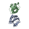 2iprSC 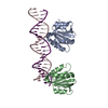 2itlC 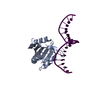 2nl8C S: Starting model for refinement C: citing same article ( |
|---|---|
| Similar structure data |


-Links
 PDBj
PDBj

- Assembly
| Deposited unit | 
| ||||||||
|---|---|---|---|---|---|---|---|---|---|
| 1 |
| ||||||||
| Unit cell |
|
-Components
| #1: Protein | Mass: 15380.704 Da / Num. of mol.: 2 / Fragment: Origin binding domain (residues 131-259) Source method: isolated from a genetically manipulated source Source: (gene. exp.) Simian virus 40 / Genus: Polyomavirus / Strain: 776 / Gene: large T antigen / Plasmid: pET15B / Species (production host): Escherichia coli / Production host:  #2: Water | ChemComp-HOH / |  Mass: 18.015 Da / Num. of mol.: 31 / Source method: isolated from a natural source / Formula: H2O Mass: 18.015 Da / Num. of mol.: 31 / Source method: isolated from a natural source / Formula: H2O |
|---|
-Experimental details
-Experiment
| Experiment | Method: X-RAY DIFFRACTION / Number of used crystals: 1 |
|---|
- Sample preparation
| Crystal | Density Matthews: 2 Å3/Da / Density % sol: 38.35 % |
|---|---|
| Crystal grow | Temperature: 297 K / Method: vapor diffusion, sitting drop / pH: 7.5 Details: 20 mM NaAc, 20% PEG4K, pH 7.5, VAPOR DIFFUSION, SITTING DROP, temperature 297K |
-Data collection
| Diffraction source | Source: ROTATING ANODE / Type: RIGAKU / Wavelength: 1.5418 Å |
|---|---|
| Detector | Type: RIGAKU RAXIS IV / Detector: IMAGE PLATE / Date: Jul 29, 2002 / Details: mirirs |
| Radiation | Monochromator: Yale mirrors / Protocol: SINGLE WAVELENGTH / Monochromatic (M) / Laue (L): M / Scattering type: x-ray |
| Radiation wavelength | Wavelength: 1.5418 Å / Relative weight: 1 |
| Reflection | Resolution: 2.5→20 Å / Num. obs: 8118 / % possible obs: 90.2 % / Observed criterion σ(I): -3 / Redundancy: 2.5 % / Rsym value: 0.088 / Net I/σ(I): 10 |
| Reflection shell | Resolution: 2.5→2.59 Å / Num. unique all: 751 / Rsym value: 0.435 / % possible all: 84.7 |
- Processing
| Software |
| |||||||||||||||||||||||||
|---|---|---|---|---|---|---|---|---|---|---|---|---|---|---|---|---|---|---|---|---|---|---|---|---|---|---|
| Refinement | Method to determine structure: MOLECULAR REPLACEMENT Starting model: 2IPR Resolution: 2.5→20 Å / Isotropic thermal model: isotropic / Cross valid method: THROUGHOUT / σ(F): 0 / Stereochemistry target values: Engh & Huber
| |||||||||||||||||||||||||
| Refinement step | Cycle: LAST / Resolution: 2.5→20 Å
| |||||||||||||||||||||||||
| Refine LS restraints |
| |||||||||||||||||||||||||
| LS refinement shell | Resolution: 2.5→2.59 Å |
