[English] 日本語
Yorodumi
- PDB-6z1d: Crystal structure of the AAA domain of Rubisco Activase from Nost... -

+
Open data


ID or keywords:

Loading...

-
Basic information

Entry
Database: PDB / ID: 6z1d
TitleCrystal structure of the AAA domain of Rubisco Activase from Nostoc sp. (strain PCC 7120), Gadolinium complex
ComponentsRibulose bisphosphate carboxylase/oxygenase activase
KeywordsCHAPERONE / AAA+ domain
Function / homology
Function and homology information


ATP hydrolysis activity / ATP binding
Similarity search - Function
Ribulose bisphosphate carboxylase/oxygenase activase-like / : / Ribulose bisphosphate carboxylase/oxygenase activase, AAA, helical / Ribulose bisphosphate carboxylase small subunit, domain / Ribulose bisphosphate carboxylase, small subunit superfamily / Ribulose bisphosphate carboxylase, small chain / Ribulose bisphosphate carboxylase, small chain / ATPase family associated with various cellular activities (AAA) / ATPase, AAA-type, core / P-loop containing nucleoside triphosphate hydrolase
Similarity search - Domain/homology
ADENOSINE-5'-DIPHOSPHATE / GADOLINIUM ATOM / Ribulose bisphosphate carboxylase/oxygenase activase
Similarity search - Component
Biological speciesNostoc sp. PCC 7120 = FACHB-418 (bacteria)
MethodX-RAY DIFFRACTION / SYNCHROTRON / MAD / Resolution: 2.705 Å
AuthorsPopilka, L. / Bracher, A.
Citation
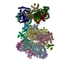
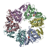
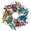
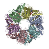
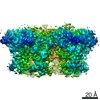
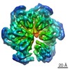
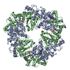
Journal: Cell / Year: 2020
Title: Dual Functions of a Rubisco Activase in Metabolic Repair and Recruitment to Carboxysomes.
Authors: Mirkko Flecken / Huping Wang / Leonhard Popilka / F Ulrich Hartl / Andreas Bracher / Manajit Hayer-Hartl /
Abstract: Rubisco, the key enzyme of CO fixation in photosynthesis, is prone to inactivation by inhibitory sugar phosphates. Inhibited Rubisco undergoes conformational repair by the hexameric AAA+ chaperone ...Rubisco, the key enzyme of CO fixation in photosynthesis, is prone to inactivation by inhibitory sugar phosphates. Inhibited Rubisco undergoes conformational repair by the hexameric AAA+ chaperone Rubisco activase (Rca) in a process that is not well understood. Here, we performed a structural and mechanistic analysis of cyanobacterial Rca, a close homolog of plant Rca. In the Rca:Rubisco complex, Rca is positioned over the Rubisco catalytic site under repair and pulls the N-terminal tail of the large Rubisco subunit (RbcL) into the hexamer pore. Simultaneous displacement of the C terminus of the adjacent RbcL opens the catalytic site for inhibitor release. An alternative interaction of Rca with Rubisco is mediated by C-terminal domains that resemble the small Rubisco subunit. These domains, together with the N-terminal AAA+ hexamer, ensure that Rca is packaged with Rubisco into carboxysomes. The cyanobacterial Rca is a dual-purpose protein with functions in Rubisco repair and carboxysome organization.
History
DepositionMay 13, 2020Deposition site: PDBE / Processing site: PDBE
Revision 1.0Sep 23, 2020Provider: repository / Type: Initial release
Revision 1.1Jan 27, 2021Group: Database references / Category: citation / citation_author
Item: _citation.journal_abbrev / _citation.journal_id_CSD ..._citation.journal_abbrev / _citation.journal_id_CSD / _citation.journal_id_ISSN / _citation.journal_volume / _citation.page_first / _citation.page_last / _citation.pdbx_database_id_DOI / _citation.pdbx_database_id_PubMed / _citation.title / _citation.year / _citation_author.identifier_ORCID

-
Structure visualization

Structure viewerMolecule:
MolmilJmol/JSmol

Downloads & links

-
Assembly

Deposited unit
A: Ribulose bisphosphate carboxylase/oxygenase activase
B: Ribulose bisphosphate carboxylase/oxygenase activase
hetero molecules


Theoretical massNumber of molelcules
Total (without water)66,6388
Polymers65,5472
Non-polymers1,0926
Water543
1
A: Ribulose bisphosphate carboxylase/oxygenase activase
B: Ribulose bisphosphate carboxylase/oxygenase activase
hetero molecules

A: Ribulose bisphosphate carboxylase/oxygenase activase
B: Ribulose bisphosphate carboxylase/oxygenase activase
hetero molecules

A: Ribulose bisphosphate carboxylase/oxygenase activase
B: Ribulose bisphosphate carboxylase/oxygenase activase
hetero molecules


Theoretical massNumber of molelcules
Total (without water)199,91524
Polymers196,6406
Non-polymers3,27518
Water1086
TypeNameSymmetry operationNumber
identity operation1_555x,y,z1
crystal symmetry operation2_445-y-1,x-y-1,z1
crystal symmetry operation3_545-x+y,-x-1,z1
Buried area24520 Å2
ΔGint-221 kcal/mol
Surface area68460 Å2
MethodPISA
Unit cell
Length a, b, c (Å)111.990, 111.990, 283.818
Angle α, β, γ (deg.)90.000, 90.000, 120.000
Int Tables number155
Space group name H-MH32

-
Components

#1: Protein Ribulose bisphosphate carboxylase/oxygenase activase / RuBisCO activase


Mass: 32773.316 Da / Num. of mol.: 2 / Fragment: AAA+ domain
Source method: isolated from a genetically manipulated source
Source: (gene. exp.) Nostoc sp. PCC 7120 = FACHB-418 (bacteria)
Gene: rca, alr1533 / Plasmid: pHUE / Production host: Escherichia coli BL21 (bacteria) / Variant (production host): pBAD33-EcGroSEL / References: UniProt: P58555
#2: Chemical
ChemComp-GD / GADOLINIUM ATOM


Mass: 157.250 Da / Num. of mol.: 4 / Source method: obtained synthetically / Formula: Gd
#3: Chemical ChemComp-CL / CHLORIDE ION / Chloride


Mass: 35.453 Da / Num. of mol.: 1 / Source method: obtained synthetically / Formula: Cl
#4: Chemical ChemComp-ADP / ADENOSINE-5'-DIPHOSPHATE / Adenosine diphosphate


Mass: 427.201 Da / Num. of mol.: 1 / Source method: obtained synthetically / Formula: C10H15N5O10P2 / Comment: ADP, energy-carrying molecule*YM
#5: Water ChemComp-HOH / water / Water


Mass: 18.015 Da / Num. of mol.: 3 / Source method: isolated from a natural source / Formula: H2O
Has ligand of interestN

-
Experimental details

-
Experiment

ExperimentMethod: X-RAY DIFFRACTION / Number of used crystals: 1

-
Sample preparation

CrystalDensity Matthews: 2.61 Å3/Da / Density % sol: 52.92 % / Mosaicity: 0 °
Crystal growTemperature: 277 K / Method: vapor diffusion / pH: 7 / Details: 2.2 M Na-acetate

-
Data collection

DiffractionMean temperature: 100 K / Serial crystal experiment: N
Diffraction sourceSource: SYNCHROTRON / Site: SLS / Beamline: X06DA / Wavelength: 1.68745 Å
DetectorType: DECTRIS PILATUS 2M / Detector: PIXEL / Date: Feb 9, 2013
RadiationProtocol: MAD / Monochromatic (M) / Laue (L): M / Scattering type: x-ray
Radiation wavelengthWavelength: 1.68745 Å / Relative weight: 1
ReflectionResolution: 2.705→48.99 Å / Num. all: 19125 / Num. obs: 19125 / % possible obs: 99.9 % / Redundancy: 19.7 % / Biso Wilson estimate: 61.79 Å2 / Rpim(I) all: 0.015 / Rrim(I) all: 0.069 / Rsym value: 0.064 / Net I/av σ(I): 11 / Net I/σ(I): 37.2 / Num. measured all: 375979
Reflection shell

Diffraction-ID: 1

Resolution (Å)Redundancy (%)Rmerge(I) obsMean I/σ(I) obsNum. unique obsRpim(I) allRrim(I) allRsym value% possible all
2.705-2.8519.30.6241.327380.1480.6580.62499.5
2.85-3.02190.3752.126270.090.3950.375100
3.02-3.2319.80.2163.624420.0510.2270.216100
3.23-3.4920.70.1365.623030.0310.1430.136100
3.49-3.8320.30.0858.821070.020.0890.085100
3.83-4.2818.90.05413.119160.0130.0580.054100
4.28-4.9420.10.03917.917070.0090.0420.039100
4.94-6.0520.30.0417.414630.010.0440.04100
6.05-8.5518.40.02922.211480.0080.0350.029100
8.55-48.9919.30.017356740.0050.0220.01799.6

-
Phasing

PhasingMethod: MAD

-
Processing

Software
NameVersionClassification
PHENIX1.8.2refinement
XDSVERSION September 26, 2012data reduction
SCALA3.3.16data scaling
SHELXDEVersion 2006/3phasing
PDB_EXTRACT3.25data extraction
RefinementMethod to determine structure: MAD / Resolution: 2.705→28.633 Å / FOM work R set: 0.8015 / SU ML: 0.39 / Cross valid method: THROUGHOUT / σ(F): 1.34 / Phase error: 26.26 / Stereochemistry target values: ML
RfactorNum. reflection% reflection
Rfree0.2585 978 5.12 %
Rwork0.2159 18124 -
obs0.2179 19102 99.9 %
Solvent computationShrinkage radii: 0.9 Å / VDW probe radii: 1.11 Å / Solvent model: FLAT BULK SOLVENT MODEL
Displacement parametersBiso max: 183.73 Å2 / Biso mean: 71.22 Å2 / Biso min: 36.87 Å2
Refinement stepCycle: final / Resolution: 2.705→28.633 Å
ProteinNucleic acidLigandSolventTotal
Num. atoms4152 0 44 3 4199
Biso mean--61.34 40.87 -
Num. residues----533
Refine LS restraints
Refine-IDTypeDev idealNumber
X-RAY DIFFRACTIONf_bond_d0.0084260
X-RAY DIFFRACTIONf_angle_d1.0985775
X-RAY DIFFRACTIONf_chiral_restr0.054654
X-RAY DIFFRACTIONf_plane_restr0.005752
X-RAY DIFFRACTIONf_dihedral_angle_d14.1621587
LS refinement shell

Refine-ID: X-RAY DIFFRACTION / Rfactor Rfree error: 0

Resolution (Å)Rfactor RfreeNum. reflection RfreeRfactor RworkNum. reflection Rwork% reflection obs (%)
2.705-2.84720.35361510.3115251399
2.8472-3.02540.33841570.26872561100
3.0254-3.25870.2821310.2642556100
3.2587-3.5860.2841350.24112575100
3.586-4.10360.24791460.21172577100
4.1036-5.16520.20181210.17792622100
5.1652-28.6330.24991370.19782720100
Refinement TLS params.

Method: refined / Refine-ID: X-RAY DIFFRACTION

IDL112)L122)L132)L222)L232)L332)S11 (Å °)S12 (Å °)S13 (Å °)S21 (Å °)S22 (Å °)S23 (Å °)S31 (Å °)S32 (Å °)S33 (Å °)T112)T122)T132)T222)T232)T332)Origin x (Å)Origin y (Å)Origin z (Å)
13.2392-1.0338-1.03214.09-0.71085.22540.00470.19110.1079-0.54350.12060.06120.1273-0.0225-0.02760.5078-0.1143-0.09280.49460.04720.4992-28.0181-44.4704-34.2397
22.8070.7099-0.54285.57411.8972.3514-0.00930.14850.0103-0.14640.00570.4258-0.123-0.28520.06430.44880.02790.04360.57780.00220.434-41.9615-68.3384-14.7184
35.00520.0181-0.37734.58550.52313.3105-0.22160.0121-0.0279-0.27030.0719-0.0264-0.0763-0.14590.14550.46990.0506-0.07580.4399-0.06540.29672.5446-34.4851-27.6098
48.61932.77924.44821.51872.18353.6314-0.1996-0.82880.32250.00680.11180.5095-0.2126-0.43440.10180.60070.1916-0.01260.8489-0.08720.7956-29.0806-27.0409-16.3893
Refinement TLS group
IDRefine-IDRefine TLS-IDSelection detailsAuth asym-IDAuth seq-ID
1X-RAY DIFFRACTION1chain 'A' and (resid 2 through 179)A2 - 179
2X-RAY DIFFRACTION2chain 'A' and (resid 180 through 275 )A180 - 275
3X-RAY DIFFRACTION3chain 'B' and (resid 2 through 179)B2 - 179
4X-RAY DIFFRACTION4chain 'B' and (resid 180 through 278)B180 - 278

+
About Yorodumi

-
News

-
Feb 9, 2022. New format data for meta-information of EMDB entries

New format data for meta-information of EMDB entries

  • Version 3 of the EMDB header file is now the official format.
  • The previous official version 1.9 will be removed from the archive.

Related info.:EMDB header

External links:wwPDB to switch to version 3 of the EMDB data model

-
Aug 12, 2020. Covid-19 info

Covid-19 info

URL: https://pdbj.org/emnavi/covid19.php

New page: Covid-19 featured information page in EM Navigator.

Related info.:Covid-19 info / Mar 5, 2020. Novel coronavirus structure data

+
Mar 5, 2020. Novel coronavirus structure data

Novel coronavirus structure data

Related info.:Yorodumi Speices / Aug 12, 2020. Covid-19 info

External links:COVID-19 featured content - PDBj / Molecule of the Month (242):Coronavirus Proteases

+
Jan 31, 2019. EMDB accession codes are about to change! (news from PDBe EMDB page)

EMDB accession codes are about to change! (news from PDBe EMDB page)

  • The allocation of 4 digits for EMDB accession codes will soon come to an end. Whilst these codes will remain in use, new EMDB accession codes will include an additional digit and will expand incrementally as the available range of codes is exhausted. The current 4-digit format prefixed with “EMD-” (i.e. EMD-XXXX) will advance to a 5-digit format (i.e. EMD-XXXXX), and so on. It is currently estimated that the 4-digit codes will be depleted around Spring 2019, at which point the 5-digit format will come into force.
  • The EM Navigator/Yorodumi systems omit the EMD- prefix.

Related info.:Q: What is EMD? / ID/Accession-code notation in Yorodumi/EM Navigator

External links:EMDB Accession Codes are Changing Soon! / Contact to PDBj

+
Jul 12, 2017. Major update of PDB

Major update of PDB

  • wwPDB released updated PDB data conforming to the new PDBx/mmCIF dictionary.
  • This is a major update changing the version number from 4 to 5, and with Remediation, in which all the entries are updated.
  • In this update, many items about electron microscopy experimental information are reorganized (e.g. em_software).
  • Now, EM Navigator and Yorodumi are based on the updated data.

External links:wwPDB Remediation / Enriched Model Files Conforming to OneDep Data Standards Now Available in the PDB FTP Archive

-
Yorodumi

Thousand views of thousand structures

  • Yorodumi is a browser for structure data from EMDB, PDB, SASBDB, etc.
  • This page is also the successor to EM Navigator detail page, and also detail information page/front-end page for Omokage search.
  • The word "yorodu" (or yorozu) is an old Japanese word meaning "ten thousand". "mi" (miru) is to see.

Related info.:EMDB / PDB / SASBDB / Comparison of 3 databanks / Yorodumi Search / Aug 31, 2016. New EM Navigator & Yorodumi / Yorodumi Papers / Jmol/JSmol / Function and homology information / Changes in new EM Navigator and Yorodumi

Read more