 Movie
Movie Controller

[English] 日本語
 Yorodumi
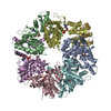
- EMDB-11789: RUVBL1-RUVBL2 heterohexameric ring after binding of RNA helicase DHX34 -
+ Open data

- Basic information
| Entry | Database: EMDB / ID: EMD-11789 | ||||||||||||
|---|---|---|---|---|---|---|---|---|---|---|---|---|---|
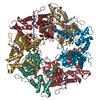
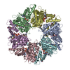
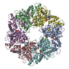
| Title | RUVBL1-RUVBL2 heterohexameric ring after binding of RNA helicase DHX34 | ||||||||||||
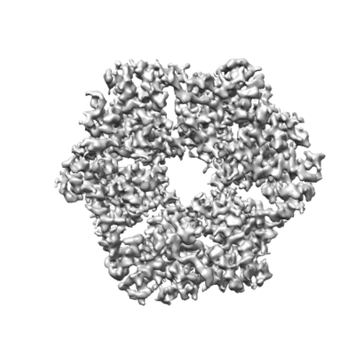
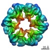
 Map data Map data | Cryo-EM structure of RUVBL1-RUVBL2 ATPase core | ||||||||||||
 Sample Sample |
| ||||||||||||
 Keywords Keywords | RUVBL1-RUVBL2 / DHX34 / Nonsense-Mediated mRNA Decay / RNA degradation / Cryo-EM / TRANSLATION | ||||||||||||
| Function / homology |  Function and homology information Function and homology informationpromoter-enhancer loop anchoring activity / telomerase RNA localization to Cajal body / establishment of protein localization to chromatin / regulation of DNA strand elongation / positive regulation of telomere maintenance in response to DNA damage / R2TP complex / dynein axonemal particle / RPAP3/R2TP/prefoldin-like complex / Swr1 complex / Ino80 complex ...promoter-enhancer loop anchoring activity / telomerase RNA localization to Cajal body / establishment of protein localization to chromatin / regulation of DNA strand elongation / positive regulation of telomere maintenance in response to DNA damage / R2TP complex / dynein axonemal particle / RPAP3/R2TP/prefoldin-like complex / Swr1 complex / Ino80 complex / regulation of double-strand break repair / box C/D snoRNP assembly / regulation of chromosome organization / NuA4 histone acetyltransferase complex / regulation of DNA replication / TFIID-class transcription factor complex binding / MLL1 complex / regulation of embryonic development / Telomere Extension By Telomerase / protein folding chaperone complex / regulation of DNA repair / RNA polymerase II core promoter sequence-specific DNA binding / positive regulation of double-strand break repair via homologous recombination / Deposition of new CENPA-containing nucleosomes at the centromere / TBP-class protein binding / telomere maintenance / positive regulation of DNA repair / DNA helicase activity / cellular response to estradiol stimulus / euchromatin / negative regulation of canonical Wnt signaling pathway / Formation of the beta-catenin:TCF transactivating complex / ADP binding / chromatin DNA binding / beta-catenin binding / DNA Damage Recognition in GG-NER / nuclear matrix / cellular response to UV / : / transcription corepressor activity / UCH proteinases / positive regulation of canonical Wnt signaling pathway / nucleosome / HATs acetylate histones / ATPase binding / protein folding / DNA recombination / spermatogenesis / regulation of apoptotic process / DNA helicase / transcription coactivator activity / regulation of cell cycle / nuclear speck / protein stabilization / Ub-specific processing proteases / ciliary basal body / RNA polymerase II cis-regulatory region sequence-specific DNA binding / cadherin binding / chromatin remodeling / ribonucleoprotein complex / cell division / DNA repair / regulation of transcription by RNA polymerase II / regulation of DNA-templated transcription / centrosome / positive regulation of DNA-templated transcription / protein homodimerization activity / ATP hydrolysis activity / positive regulation of transcription by RNA polymerase II / extracellular exosome / nucleoplasm / ATP binding / membrane / identical protein binding / nucleus / cytoplasm / cytosol Similarity search - Function | ||||||||||||
| Biological species |  Homo sapiens (human) Homo sapiens (human) | ||||||||||||
| Method | single particle reconstruction / cryo EM / Resolution: 4.18 Å | ||||||||||||
 Authors Authors | Lopez-Perrote A / Rodriguez CF / Llorca O | ||||||||||||
| Funding support |  Spain, 3 items Spain, 3 items
| ||||||||||||
 Citation Citation | Journal: Elife / Year: 2020 Title: Regulation of RUVBL1-RUVBL2 AAA-ATPases by the nonsense-mediated mRNA decay factor DHX34, as evidenced by Cryo-EM. Authors: Andres López-Perrote / Nele Hug / Ana González-Corpas / Carlos F Rodríguez / Marina Serna / Carmen García-Martín / Jasminka Boskovic / Rafael Fernandez-Leiro / Javier F Caceres / Oscar Llorca /  Abstract: Nonsense-mediated mRNA decay (NMD) is a surveillance pathway that degrades aberrant mRNAs and also regulates the expression of a wide range of physiological transcripts. RUVBL1 and RUVBL2 AAA-ATPases ...Nonsense-mediated mRNA decay (NMD) is a surveillance pathway that degrades aberrant mRNAs and also regulates the expression of a wide range of physiological transcripts. RUVBL1 and RUVBL2 AAA-ATPases form an hetero-hexameric ring that is part of several macromolecular complexes such as INO80, SWR1, and R2TP. Interestingly, RUVBL1-RUVBL2 ATPase activity is required for NMD activation by an unknown mechanism. Here, we show that DHX34, an RNA helicase regulating NMD initiation, directly interacts with RUVBL1-RUVBL2 in vitro and in cells. Cryo-EM reveals that DHX34 induces extensive changes in the N-termini of every RUVBL2 subunit in the complex, stabilizing a conformation that does not bind nucleotide and thereby down-regulates ATP hydrolysis of the complex. Using ATPase-deficient mutants, we find that DHX34 acts exclusively on the RUVBL2 subunits. We propose a model, where DHX34 acts to couple RUVBL1-RUVBL2 ATPase activity to the assembly of factors required to initiate the NMD response. | ||||||||||||
| History |
|
- Structure visualization
| Movie |
Movie viewer |
|---|---|
| Structure viewer | EM map: SurfViewMolmilJmol/JSmol |
| Supplemental images |
- Downloads & links
-EMDB archive
| Map data | emd_11789.map.gz | 5.9 MB | EMDB map data format | |
|---|---|---|---|---|
| Header (meta data) | emd-11789-v30.xmlemd-11789.xml | 21.3 KB 21.3 KB | Display Display | EMDB header |
| FSC (resolution estimation) | emd_11789_fsc.xml | 10 KB | Display | FSC data file |
| Images |  emd_11789.png emd_11789.png | 132.9 KB | ||
| Filedesc metadata | emd-11789.cif.gz | 7 KB | ||
| Archive directory |  http://ftp.pdbj.org/pub/emdb/structures/EMD-11789ftp://ftp.pdbj.org/pub/emdb/structures/EMD-11789 http://ftp.pdbj.org/pub/emdb/structures/EMD-11789ftp://ftp.pdbj.org/pub/emdb/structures/EMD-11789 | HTTPS FTP |
-Related structure data
| Related structure data |  7ahoMC C: citing same article ( M: atomic model generated by this map |
|---|---|
| Similar structure data |


-Links
| EMDB pages | EMDB (EBI/PDBe) / EMDataResource |
|---|---|
| Related items in Molecule of the Month |


-Map
| File | Download / File: emd_11789.map.gz / Format: CCP4 / Size: 83.7 MB / Type: IMAGE STORED AS FLOATING POINT NUMBER (4 BYTES) | ||||||||||||||||||||||||||||||||||||||||||||||||||||||||||||
|---|---|---|---|---|---|---|---|---|---|---|---|---|---|---|---|---|---|---|---|---|---|---|---|---|---|---|---|---|---|---|---|---|---|---|---|---|---|---|---|---|---|---|---|---|---|---|---|---|---|---|---|---|---|---|---|---|---|---|---|---|---|
| Annotation | Cryo-EM structure of RUVBL1-RUVBL2 ATPase core | ||||||||||||||||||||||||||||||||||||||||||||||||||||||||||||
| Projections & slices | Image control
Images are generated by Spider. | ||||||||||||||||||||||||||||||||||||||||||||||||||||||||||||
| Voxel size | X=Y=Z: 1.047 Å | ||||||||||||||||||||||||||||||||||||||||||||||||||||||||||||
| Density |
| ||||||||||||||||||||||||||||||||||||||||||||||||||||||||||||
| Symmetry | Space group: 1 | ||||||||||||||||||||||||||||||||||||||||||||||||||||||||||||
| Details | EMDB XML:
CCP4 map header:
| ||||||||||||||||||||||||||||||||||||||||||||||||||||||||||||
 Z (Sec.)
 Y (Row.)
 X (Col.)


-Supplemental data
- Sample components
-Entire : RUVBL1-RUVBL2 heterohexameric ring after binding of RNA helicase DHX34
| Entire | Name: RUVBL1-RUVBL2 heterohexameric ring after binding of RNA helicase DHX34 |
|---|---|
| Components |
|
-Supramolecule #1: RUVBL1-RUVBL2 heterohexameric ring after binding of RNA helicase DHX34
| Supramolecule | Name: RUVBL1-RUVBL2 heterohexameric ring after binding of RNA helicase DHX34 type: complex / ID: 1 / Parent: 0 / Macromolecule list: #1-#2 Details: Structure of the RUVBL1-RUVBL2 hetero-hexameric ring region after the interaction of RNA helicase DHX34 |
|---|---|
| Source (natural) | Organism: Homo sapiens (human) |
| Molecular weight | Theoretical: 0.44928 kDa/nm |
-Macromolecule #1: RuvB-like 1
| Macromolecule | Name: RuvB-like 1 / type: protein_or_peptide / ID: 1 / Number of copies: 3 / Enantiomer: LEVO / EC number: DNA helicase |
|---|---|
| Source (natural) | Organism: Homo sapiens (human) |
| Molecular weight | Theoretical: 52.710406 KDa |
| Recombinant expression | Organism:  |
| Sequence | String: HHHHHHHHSS GENLYFQGSH MKIEEVKSTT KTQRIASHSH VKGLGLDESG LAKQAASGLV GQENAREACG VIVELIKSKK MAGRAVLLA GPPGTGKTAL ALAIAQELGS KVPFCPMVGS EVYSTEIKKT EVLMENFRRA IGLRIKETKE VYEGEVTELT P CETENPMG ...String: HHHHHHHHSS GENLYFQGSH MKIEEVKSTT KTQRIASHSH VKGLGLDESG LAKQAASGLV GQENAREACG VIVELIKSKK MAGRAVLLA GPPGTGKTAL ALAIAQELGS KVPFCPMVGS EVYSTEIKKT EVLMENFRRA IGLRIKETKE VYEGEVTELT P CETENPMG GYGKTISHVI IGLKTAKGTK QLKLDPSIFE SLQKERVEAG DVIYIEANSG AVKRQGRCDT YATEFDLEAE EY VPLPKGD VHKKKEIIQD VTLHDLDVAN ARPQGGQDIL SMMGQLMKPK KTEITDKLRG EINKVVNKYI DQGIAELVPG VLF VDEVHM LDIECFTYLH RALESSIAPI VIFASNRGNC VIRGTEDITS PHGIPLDLLD RVMIIRTMLY TPQEMKQIIK IRAQ TEGIN ISEEALNHLG EIGTKTTLRY SVQLLTPANL LAKINGKDSI EKEHVEEISE LFYDAKSSAK ILADQQDKYM K UniProtKB: RuvB-like 1 |
-Macromolecule #2: RuvB-like 2
| Macromolecule | Name: RuvB-like 2 / type: protein_or_peptide / ID: 2 / Number of copies: 3 / Enantiomer: LEVO / EC number: DNA helicase |
|---|---|
| Source (natural) | Organism: Homo sapiens (human) |
| Molecular weight | Theoretical: 53.047488 KDa |
| Recombinant expression | Organism: |
| Sequence | String: MADLNWISAG HAIADVGTMA TVTATTKVPE IRDVTRIERI GAHSHIRGLG LDDALEPRQA SQGMVGQLAA RRAAGVVLEM IREGKIAGR AVLIAGQPGT GKTAIAMGMA QALGPDTPFT AIAGSEIFSL EMSKTEALTQ AFRRSIGVRI KEETEIIEGE V VEIQIDRP ...String: MADLNWISAG HAIADVGTMA TVTATTKVPE IRDVTRIERI GAHSHIRGLG LDDALEPRQA SQGMVGQLAA RRAAGVVLEM IREGKIAGR AVLIAGQPGT GKTAIAMGMA QALGPDTPFT AIAGSEIFSL EMSKTEALTQ AFRRSIGVRI KEETEIIEGE V VEIQIDRP ATGTGSKVGK LTLKTTEMET IYDLGTKMIE SLTKDKVQAG DVITIDKATG KISKLGRSFT RARDYDAMGS QT KFVQCPD GELQKRKEVV HTVSLHEIDV INSRTQGFLA LFSGDTGEIK SEVREQINAK VAEWREEGKA EIIPGVLFID EVH MLDIES FSFLNRALES DMAPVLIMAT NRGITRIRGT SYQSPHGIPI DLLDRLLIVS TTPYSEKDTK QILRIRCEEE DVEM SEDAY TVLTRIGLET SLRYAIQLIT AASLVCRKRK GTEVQVDDIK RVYSLFLDES RSTQYMKEYQ DAFLFNELKG ETMDT S UniProtKB: RuvB-like 2 |
-Macromolecule #3: ADENOSINE-5'-DIPHOSPHATE
| Macromolecule | Name: ADENOSINE-5'-DIPHOSPHATE / type: ligand / ID: 3 / Number of copies: 3 / Formula: ADP |
|---|---|
| Molecular weight | Theoretical: 427.201 Da |
| Chemical component information |  ChemComp-ADP: |
-Experimental details
-Structure determination
| Method | cryo EM |
|---|---|
 Processing Processing | single particle reconstruction |
| Aggregation state | particle |
-Sample preparation
| Concentration | 0.35 mg/mL |
|---|---|
| Buffer | pH: 7.4 / Component - Formula: Tris-NaCl / Component - Name: TBS / Details: 50 mM Tris-HCl pH 7.4, 150 mM NaCl |
| Vitrification | Cryogen name: ETHANE / Chamber humidity: 95 % / Chamber temperature: 277.15 K / Instrument: FEI VITROBOT MARK IV |
- Electron microscopy
| Microscope | FEI TITAN KRIOS |
|---|---|
| Image recording | Film or detector model: GATAN K2 SUMMIT (4k x 4k) / Detector mode: COUNTING / Number grids imaged: 1 / Number real images: 3047 / Average electron dose: 48.1 e/Å2 |
| Electron beam | Acceleration voltage: 300 kV / Electron source:  FIELD EMISSION GUN FIELD EMISSION GUN |
| Electron optics | Calibrated magnification: 47756 / Illumination mode: FLOOD BEAM / Imaging mode: BRIGHT FIELD / Cs: 2.7 mm / Nominal defocus max: 0.003 µm / Nominal defocus min: 0.0015 µm |
| Sample stage | Specimen holder model: FEI TITAN KRIOS AUTOGRID HOLDER / Cooling holder cryogen: NITROGEN |
| Experimental equipment |  Model: Titan Krios / Image courtesy: FEI Company |


