 Movie
Movie Controller

[English] 日本語
 Yorodumi
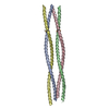
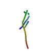
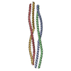
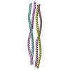
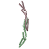
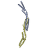
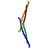
- PDB-6yrn: Structure of the Chlamydomonas reinhardtii SAS-6 coiled-coil doma... -
+ Open data

- Basic information
| Entry | Database: PDB / ID: 6yrn | |||||||||
|---|---|---|---|---|---|---|---|---|---|---|
| Title | Structure of the Chlamydomonas reinhardtii SAS-6 coiled-coil domain, P2 crystal form | |||||||||
 Components Components | Centriole protein | |||||||||
 Keywords Keywords | STRUCTURAL PROTEIN / centriole / centrosome / cartwheel / coiled coil / complex / alpha helical | |||||||||
| Function / homology | : / Sas-6-like, oligomerization domain / Spindle assembly abnormal protein 6, N-terminal / SAS-6, N-terminal domain superfamily / Centriolar protein SAS N-terminal domain / identical protein binding / Centriole protein Function and homology information Function and homology information | |||||||||
| Biological species |   Chlamydomonas reinhardtii (plant) Chlamydomonas reinhardtii (plant) | |||||||||
| Method |  X-RAY DIFFRACTION / SYNCHROTRON / MOLECULAR REPLACEMENT / Resolution: 2.43 Å X-RAY DIFFRACTION / SYNCHROTRON / MOLECULAR REPLACEMENT / Resolution: 2.43 Å | |||||||||
 Authors Authors | Kantsadi, A.L. / Vakonakis, I. | |||||||||
| Funding support |  United Kingdom, 2items United Kingdom, 2items
| |||||||||
 Citation Citation | Journal: Structure / Year: 2022 Title: Structures of SAS-6 coiled coil hold implications for the polarity of the centriolar cartwheel. Authors: Kantsadi, A.L. / Hatzopoulos, G.N. / Gonczy, P. / Vakonakis, I. | |||||||||
| History |
|
- Structure visualization
| Structure viewer | Molecule: MolmilJmol/JSmol |
|---|
- Downloads & links
-Download
| PDBx/mmCIF format | 6yrn.cif.gz | 333.9 KB | Display | PDBx/mmCIF format |
|---|---|---|---|---|
| PDB format | pdb6yrn.ent.gz | 276.4 KB | Display | PDB format |
| PDBx/mmJSON format | 6yrn.json.gz | Tree view | PDBx/mmJSON format | |
| Others |  Other downloads Other downloads |
-Validation report
| Arichive directory | https://data.pdbj.org/pub/pdb/validation_reports/yr/6yrnftp://data.pdbj.org/pub/pdb/validation_reports/yr/6yrn | HTTPS FTP |
|---|
-Related structure data
| Related structure data |  6yrlSC 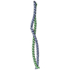 6ys4C  6z26C S: Starting model for refinement C: citing same article ( |
|---|---|
| Similar structure data |
-Links
 PDBj
PDBj- Assembly
| Deposited unit | 
| ||||||||
|---|---|---|---|---|---|---|---|---|---|
| 1 | 
| ||||||||
| 2 | 
| ||||||||
| Unit cell |
|
-Components
| #1: Protein | Mass: 13010.449 Da / Num. of mol.: 8 Source method: isolated from a genetically manipulated source Source: (gene. exp.) Chlamydomonas reinhardtii (plant) / Gene: CrSAS-6, CHLRE_12g516950v5 / Plasmid: pFLOATDetails (production host): pET-30a derivative, N-terminal His6-tag Production host:  #2: Chemical | ChemComp-PG4 / |   Mass: 194.226 Da / Num. of mol.: 1 / Source method: obtained synthetically / Formula: C8H18O5 / Comment: precipitant*YM Mass: 194.226 Da / Num. of mol.: 1 / Source method: obtained synthetically / Formula: C8H18O5 / Comment: precipitant*YM#3: Water | ChemComp-HOH / |  Mass: 18.015 Da / Num. of mol.: 69 / Source method: isolated from a natural source / Formula: H2O Mass: 18.015 Da / Num. of mol.: 69 / Source method: isolated from a natural source / Formula: H2OHas ligand of interest | N | |
|---|
-Experimental details
-Experiment
| Experiment | Method: X-RAY DIFFRACTION / Number of used crystals: 1 |
|---|
- Sample preparation
| Crystal | Density Matthews: 2.56 Å3/Da / Density % sol: 51.94 % |
|---|---|
| Crystal grow | Temperature: 277 K / Method: vapor diffusion, sitting drop / pH: 6.5 Details: 0.1 M carboxylic acid mixture, 0.1 M imidazole / MES buffer 20% v/v ethylene glycol 10% w/v PEG 8000 |
-Data collection
| Diffraction | Mean temperature: 100 K / Serial crystal experiment: N |
|---|---|
| Diffraction source | Source: SYNCHROTRON / Site: Diamond / Beamline: I04 / Wavelength: 0.9795 Å |
| Detector | Type: DECTRIS PILATUS 6M-F / Detector: PIXEL / Date: Jul 23, 2017 |
| Radiation | Protocol: SINGLE WAVELENGTH / Monochromatic (M) / Laue (L): M / Scattering type: x-ray |
| Radiation wavelength | Wavelength: 0.9795 Å / Relative weight: 1 |
| Reflection | Resolution: 2.43→84.82 Å / Num. obs: 40623 / % possible obs: 99.8 % / Redundancy: 3.2 % / Biso Wilson estimate: 37.41 Å2 / CC1/2: 0.995 / Rmerge(I) obs: 0.154 / Rpim(I) all: 0.101 / Rrim(I) all: 0.185 / Net I/σ(I): 3.7 |
| Reflection shell | Resolution: 2.43→2.47 Å / Redundancy: 2.9 % / Rmerge(I) obs: 0.855 / Mean I/σ(I) obs: 1.1 / Num. unique obs: 2015 / CC1/2: 0.492 / Rpim(I) all: 0.601 / Rrim(I) all: 1.065 / % possible all: 98.9 |
- Processing
| Software |
| |||||||||||||||||||||||||||||||||||||||||||||||||||||||||||||||||||||||||||||||||||||||||||||||||||||||||||||||||||||||||||||||||||||||||||||||||||||||||||||||||||||||||||||||||||||||||||||||||||||||||||||||||||||||||||||||||
|---|---|---|---|---|---|---|---|---|---|---|---|---|---|---|---|---|---|---|---|---|---|---|---|---|---|---|---|---|---|---|---|---|---|---|---|---|---|---|---|---|---|---|---|---|---|---|---|---|---|---|---|---|---|---|---|---|---|---|---|---|---|---|---|---|---|---|---|---|---|---|---|---|---|---|---|---|---|---|---|---|---|---|---|---|---|---|---|---|---|---|---|---|---|---|---|---|---|---|---|---|---|---|---|---|---|---|---|---|---|---|---|---|---|---|---|---|---|---|---|---|---|---|---|---|---|---|---|---|---|---|---|---|---|---|---|---|---|---|---|---|---|---|---|---|---|---|---|---|---|---|---|---|---|---|---|---|---|---|---|---|---|---|---|---|---|---|---|---|---|---|---|---|---|---|---|---|---|---|---|---|---|---|---|---|---|---|---|---|---|---|---|---|---|---|---|---|---|---|---|---|---|---|---|---|---|---|---|---|---|---|---|---|---|---|---|---|---|---|---|---|---|---|---|---|---|---|
| Refinement | Method to determine structure: MOLECULAR REPLACEMENT Starting model: 6YRL Resolution: 2.43→81.53 Å / Cor.coef. Fo:Fc: 0.898 / Cor.coef. Fo:Fc free: 0.9 / SU R Cruickshank DPI: 0.474 / Cross valid method: THROUGHOUT / σ(F): 0 / SU R Blow DPI: 0.462 / SU Rfree Blow DPI: 0.297 / SU Rfree Cruickshank DPI: 0.304
| |||||||||||||||||||||||||||||||||||||||||||||||||||||||||||||||||||||||||||||||||||||||||||||||||||||||||||||||||||||||||||||||||||||||||||||||||||||||||||||||||||||||||||||||||||||||||||||||||||||||||||||||||||||||||||||||||
| Displacement parameters | Biso mean: 49.61 Å2
| |||||||||||||||||||||||||||||||||||||||||||||||||||||||||||||||||||||||||||||||||||||||||||||||||||||||||||||||||||||||||||||||||||||||||||||||||||||||||||||||||||||||||||||||||||||||||||||||||||||||||||||||||||||||||||||||||
| Refine analyze | Luzzati coordinate error obs: 0.5 Å | |||||||||||||||||||||||||||||||||||||||||||||||||||||||||||||||||||||||||||||||||||||||||||||||||||||||||||||||||||||||||||||||||||||||||||||||||||||||||||||||||||||||||||||||||||||||||||||||||||||||||||||||||||||||||||||||||
| Refinement step | Cycle: 1 / Resolution: 2.43→81.53 Å
| |||||||||||||||||||||||||||||||||||||||||||||||||||||||||||||||||||||||||||||||||||||||||||||||||||||||||||||||||||||||||||||||||||||||||||||||||||||||||||||||||||||||||||||||||||||||||||||||||||||||||||||||||||||||||||||||||
| Refine LS restraints |
| |||||||||||||||||||||||||||||||||||||||||||||||||||||||||||||||||||||||||||||||||||||||||||||||||||||||||||||||||||||||||||||||||||||||||||||||||||||||||||||||||||||||||||||||||||||||||||||||||||||||||||||||||||||||||||||||||
| LS refinement shell | Resolution: 2.43→2.45 Å / Total num. of bins used: 50
| |||||||||||||||||||||||||||||||||||||||||||||||||||||||||||||||||||||||||||||||||||||||||||||||||||||||||||||||||||||||||||||||||||||||||||||||||||||||||||||||||||||||||||||||||||||||||||||||||||||||||||||||||||||||||||||||||
| Refinement TLS params. | Method: refined / Refine-ID: X-RAY DIFFRACTION
| |||||||||||||||||||||||||||||||||||||||||||||||||||||||||||||||||||||||||||||||||||||||||||||||||||||||||||||||||||||||||||||||||||||||||||||||||||||||||||||||||||||||||||||||||||||||||||||||||||||||||||||||||||||||||||||||||
| Refinement TLS group |
|
