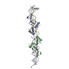
Entry Database : PDB / ID : 4x5uTitle X-ray crystal structure of CagL at pH 4.2 Cag pathogenicity island protein (Cag18) Keywords / / / Function / homology / / / / / / Biological species Helicobacter pylori (bacteria)Method / / / Resolution : 2.3 Å Authors Sundberg, E.J. / Bonsor, D.A. / Diederichs, K. Journal : J.Biol.Chem. / Year : 2015Title : Integrin Engagement by the Helical RGD Motif of the Helicobacter pylori CagL Protein Is Regulated by pH-induced Displacement of a Neighboring Helix.Authors : Bonsor, D.A. / Pham, K.T. / Beadenkopf, R. / Diederichs, K. / Haas, R. / Beckett, D. / Fischer, W. / Sundberg, E.J. History Deposition Dec 5, 2014 Deposition site / Processing site Revision 1.0 Apr 8, 2015 Provider / Type Revision 1.1 Apr 15, 2015 Group Revision 1.2 May 27, 2015 Group Revision 1.3 Nov 22, 2017 Group Database references / Derived calculations ... Database references / Derived calculations / Refinement description / Source and taxonomy Category citation / entity_src_gen ... citation / entity_src_gen / pdbx_struct_oper_list / software Item _citation.journal_id_CSD / _entity_src_gen.pdbx_alt_source_flag ... _citation.journal_id_CSD / _entity_src_gen.pdbx_alt_source_flag / _pdbx_struct_oper_list.symmetry_operation / _software.classification Revision 1.4 Sep 27, 2023 Group Data collection / Database references ... Data collection / Database references / Derived calculations / Refinement description Category chem_comp_atom / chem_comp_bond ... chem_comp_atom / chem_comp_bond / database_2 / pdbx_initial_refinement_model / pdbx_struct_special_symmetry Item / _database_2.pdbx_database_accessionRevision 1.5 Oct 16, 2024 Group / Category / pdbx_modification_feature
Show all Show less
 Movie
Movie Controller


 Open data
 Basic information
 Components
 Keywords
 Function and homology information
 Helicobacter pylori (bacteria)
 X-RAY DIFFRACTION /
  Authors
 Citation
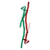
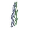
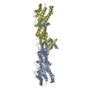
 Structure visualization
 Downloads & links
 Other downloads
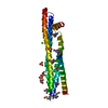


 PDBj
PDBj

 Assembly

 Mass: 18.015 Da / Num. of mol.: 3 / Source method: isolated from a natural source / Formula: H2O
 Sample preparation
 / Beamline: BL11-1 / Wavelength: 0.97945 Å
 Processing