| 登録情報 | データベース: PDB / ID: 6yn0
|
|---|
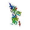
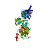
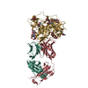
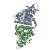
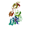
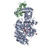
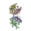
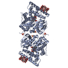
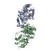
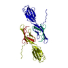
| タイトル | Structure of E. coli PBP1b with a FtsN peptide activating transglycosylase activity |
|---|
 要素 要素 | - Cell division protein FtsN
- Penicillin-binding protein 1B
|
|---|
 キーワード キーワード | TRANSFERASE / glycosyl transferase / penicillin binding protein / peptidoglycan synthesis |
|---|
| 機能・相同性 |  機能・相同性情報 機能・相同性情報
positive regulation of bipolar cell growth / cell wall repair / division septum / peptidoglycan glycosyltransferase / divisome complex / peptidoglycan glycosyltransferase activity / cell septum / peptidoglycan binding / serine-type D-Ala-D-Ala carboxypeptidase / serine-type D-Ala-D-Ala carboxypeptidase activity ...positive regulation of bipolar cell growth / cell wall repair / division septum / peptidoglycan glycosyltransferase / divisome complex / peptidoglycan glycosyltransferase activity / cell septum / peptidoglycan binding / serine-type D-Ala-D-Ala carboxypeptidase / serine-type D-Ala-D-Ala carboxypeptidase activity / division septum assembly / FtsZ-dependent cytokinesis / cell division site / penicillin binding / peptidoglycan biosynthetic process / peptidoglycan-based cell wall / regulation of cell shape / outer membrane-bounded periplasmic space / cell division / response to antibiotic / proteolysis / membrane / plasma membrane類似検索 - 分子機能 Cell division protein FtsN / : / Sporulation-like domain / Sporulation-like domain superfamily / SPOR domain / SPOR domain profile. / Penicillin-binding protein 1B / Bifunctional transglycosylase second domain / Transglycosylase PBP1b, N-terminal transmembrane domain / Transmembrane domain of transglycosylase PBP1 at N-terminal ...Cell division protein FtsN / : / Sporulation-like domain / Sporulation-like domain superfamily / SPOR domain / SPOR domain profile. / Penicillin-binding protein 1B / Bifunctional transglycosylase second domain / Transglycosylase PBP1b, N-terminal transmembrane domain / Transmembrane domain of transglycosylase PBP1 at N-terminal / Bifunctional transglycosylase second domain / Glycosyl transferase, family 51 / Penicillin binding protein transglycosylase domain / : / Transglycosylase / Penicillin-binding protein, transpeptidase / Penicillin binding protein transpeptidase domain / Beta-lactamase/transpeptidase-like / Lysozyme-like domain superfamily類似検索 - ドメイン・相同性 MOENOMYCIN / Penicillin-binding protein 1B / Cell division protein FtsN類似検索 - 構成要素 |
|---|
| 生物種 |   Escherichia coli (大腸菌) Escherichia coli (大腸菌) |
|---|
| 手法 |  X線回折 / シンクロトロン / 分子置換 / 解像度: 2.4 Å X線回折 / シンクロトロン / 分子置換 / 解像度: 2.4 Å |
|---|
 データ登録者 データ登録者 | Kerff, F. / Terrak, M. / Boes, A. / Herman, H. / Charlier, P. |
|---|
 引用 引用 | ジャーナル: J.Biol.Chem. / 年: 2020
タイトル: The bacterial cell division protein fragment E FtsN binds to and activates the major peptidoglycan synthase PBP1b.
著者: Boes, A. / Kerff, F. / Herman, R. / Touze, T. / Breukink, E. / Terrak, M. |
|---|
| 履歴 | | 登録 | 2020年4月10日 | 登録サイト: PDBE / 処理サイト: PDBE |
|---|
| 改定 1.0 | 2020年11月4日 | Provider: repository / タイプ: Initial release |
|---|
| 改定 1.1 | 2020年11月11日 | Group: Database references / カテゴリ: citation / citation_author
Item: _citation.pdbx_database_id_DOI / _citation.pdbx_database_id_PubMed ..._citation.pdbx_database_id_DOI / _citation.pdbx_database_id_PubMed / _citation.title / _citation_author.identifier_ORCID |
|---|
| 改定 1.2 | 2021年1月6日 | Group: Database references / カテゴリ: citation / citation_author
Item: _citation.journal_volume / _citation.page_first ..._citation.journal_volume / _citation.page_first / _citation.page_last / _citation_author.identifier_ORCID |
|---|
| 改定 1.3 | 2024年1月24日 | Group: Data collection / Database references / Refinement description
カテゴリ: chem_comp_atom / chem_comp_bond ...chem_comp_atom / chem_comp_bond / database_2 / pdbx_initial_refinement_model
Item: _database_2.pdbx_DOI / _database_2.pdbx_database_accession |
|---|
|
|---|
 ムービー
ムービー コントローラー


 データを開く
 基本情報
基本情報 構造の表示
構造の表示 ダウンロードとリンク
 その他のダウンロード
 PDBj
PDBj
 集合体
集合体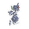
 分子量: 1580.567 Da / 分子数: 1 / 由来タイプ: 合成 / 式: C69H106N5O34P / コメント: 抗生剤*YM
 分子量: 18.015 Da / 分子数: 55 / 由来タイプ: 天然 / 式: H2O
 試料調製
試料調製 / ビームライン: PROXIMA 1 / 波長: 0.978565 Å
 解析
解析