 Movie
Movie Controller

[English] 日本語
 Yorodumi
- PDB-2jkz: SACCHAROMYCES CEREVISIAE HYPOXANTHINE-GUANINE PHOSPHORIBOSYLTRANS... -
+ Open data

- Basic information
| Entry | Database: PDB / ID: 2jkz | ||||||
|---|---|---|---|---|---|---|---|
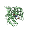
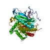
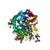
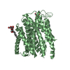
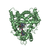
| Title | SACCHAROMYCES CEREVISIAE HYPOXANTHINE-GUANINE PHOSPHORIBOSYLTRANSFERASE IN COMPLEX WITH GMP (GUANOSINE 5'- MONOPHOSPHATE) (ORTHORHOMBIC CRYSTAL FORM) | ||||||
 Components Components | HYPOXANTHINE-GUANINE PHOSPHORIBOSYLTRANSFERASE | ||||||
 Keywords Keywords | TRANSFERASE / NUCLEUS / CYTOPLASM / MAGNESIUM / GMP COMPLEX / FLIP PEPTIDE-PLANE / GLYCOSYLTRANSFERASE / METAL-BINDING / PURINE SALVAGE | ||||||
| Function / homology |  Function and homology information Function and homology informationXMP salvage / hypoxanthine phosphoribosyltransferase / guanine phosphoribosyltransferase activity / hypoxanthine metabolic process / hypoxanthine phosphoribosyltransferase activity / GMP salvage / IMP salvage / purine ribonucleoside salvage / nucleotide binding / metal ion binding ...XMP salvage / hypoxanthine phosphoribosyltransferase / guanine phosphoribosyltransferase activity / hypoxanthine metabolic process / hypoxanthine phosphoribosyltransferase activity / GMP salvage / IMP salvage / purine ribonucleoside salvage / nucleotide binding / metal ion binding / nucleus / cytoplasm Similarity search - Function | ||||||
| Biological species |  | ||||||
| Method |  X-RAY DIFFRACTION / SYNCHROTRON / MOLECULAR REPLACEMENT / Resolution: 3.45 Å X-RAY DIFFRACTION / SYNCHROTRON / MOLECULAR REPLACEMENT / Resolution: 3.45 Å | ||||||
 Authors Authors | Moynie, L. / Giraud, M.F. / Breton, A. / Boissier, F. / Daignan-Fornier, B. / Dautant, A. | ||||||
 Citation Citation | Journal: Protein Sci. / Year: 2012 Title: Functional Significance of Four Successive Glycine Residues in the Pyrophosphate Binding Loop of Fungal 6-Oxopurine Phosphoribosyltransferases. Authors: Moynie, L. / Giraud, M.F. / Breton, A. / Boissier, F. / Daignan-Fornier, B. / Dautant, A. #1: Journal: Genetics / Year: 2008 Title: Lethal Accumulation of Guanylic Nucleotides in Saccharomyces Cerevisiae Hpt1-Deregulated Mutants. Authors: Breton, A. / Pinson, B. / Coulpier, F. / Giraud, M. / Dautant, A. / Daignan-Fornier, B. #2: Journal: J.Mol.Biol. / Year: 1998Title: Structures of Free and Complexed Forms of Escherichia Coli Xanthine-Guanine Phosphoribosyltransferase. Authors: Vos, S. / Parry, R.J. / Burns, M.R. / De Jersey, J. / Martin, J.L. #3: Journal: Biochemistry / Year: 1997Title: Crystal Structure of Escherichia Coli Xanthine Phosphoribosyltransferase. Authors: Vos, S. / De Jersey, J. / Martin, J.L. | ||||||
| History |
| ||||||
| Remark 700 | SHEET THE SHEET STRUCTURE OF THIS MOLECULE IS BIFURCATED. IN ORDER TO REPRESENT THIS FEATURE IN ... SHEET THE SHEET STRUCTURE OF THIS MOLECULE IS BIFURCATED. IN ORDER TO REPRESENT THIS FEATURE IN THE SHEET RECORDS BELOW, TWO SHEETS ARE DEFINED. |
- Structure visualization
| Structure viewer | Molecule: MolmilJmol/JSmol |
|---|
- Downloads & links
-Download
| PDBx/mmCIF format | 2jkz.cif.gz | 174.5 KB | Display | PDBx/mmCIF format |
|---|---|---|---|---|
| PDB format | pdb2jkz.ent.gz | 139 KB | Display | PDB format |
| PDBx/mmJSON format | 2jkz.json.gz | Tree view | PDBx/mmJSON format | |
| Others |  Other downloads Other downloads |
-Validation report
| Arichive directory | https://data.pdbj.org/pub/pdb/validation_reports/jk/2jkzftp://data.pdbj.org/pub/pdb/validation_reports/jk/2jkz | HTTPS FTP |
|---|
-Related structure data
| Related structure data |  2jkySC  2xbuC S: Starting model for refinement C: citing same article ( |
|---|---|
| Similar structure data |

-Links
 PDBj
PDBj

- Assembly
| Deposited unit | 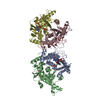
| ||||||||||||||||
|---|---|---|---|---|---|---|---|---|---|---|---|---|---|---|---|---|---|
| 1 | 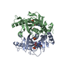
| ||||||||||||||||
| 2 | 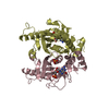
| ||||||||||||||||
| Unit cell |
| ||||||||||||||||
| Noncrystallographic symmetry (NCS) | NCS oper:
|
-Components
| #1: Protein | Mass: 25092.520 Da / Num. of mol.: 4 / Fragment: RESIDUES 2-221 Source method: isolated from a genetically manipulated source Source: (gene. exp.) Strain: Y1846 / Plasmid: PET-21 / Production host:  References: UniProt: Q04178, hypoxanthine phosphoribosyltransferase #2: Chemical | ChemComp-5GP /   Mass: 363.221 Da / Num. of mol.: 4 / Source method: obtained synthetically / Formula: C10H14N5O8P Mass: 363.221 Da / Num. of mol.: 4 / Source method: obtained synthetically / Formula: C10H14N5O8P#3: Chemical | ChemComp-SO4 /   Mass: 96.063 Da / Num. of mol.: 4 / Source method: obtained synthetically / Formula: SO4 Mass: 96.063 Da / Num. of mol.: 4 / Source method: obtained synthetically / Formula: SO4 |
|---|
-Experimental details
-Experiment
| Experiment | Method: X-RAY DIFFRACTION / Number of used crystals: 1 |
|---|
- Sample preparation
| Crystal | Density Matthews: 4.73 Å3/Da / Density % sol: 74 % / Description: NONE |
|---|---|
| Crystal grow | Temperature: 293 K / Method: vapor diffusion, sitting drop / pH: 5.8 Details: 1.6 M AMMONIUM SULPHATE, 4% PEG 400, 0.2 M NA-K TARTRATE, 50 MM NA CITRATE, 50 MM MES, PH 5.8, VAPOR DIFFUSION, SITTING DROP, TEMPERATURE 293K |
-Data collection
| Diffraction | Mean temperature: 100 K |
|---|---|
| Diffraction source | Source: SYNCHROTRON / Site: ESRF  / Beamline: BM30A / Wavelength: 0.98 / Beamline: BM30A / Wavelength: 0.98 |
| Detector | Type: MARRESEARCH / Detector: CCD / Date: Sep 10, 2005 |
| Radiation | Protocol: SINGLE WAVELENGTH / Monochromatic (M) / Laue (L): M / Scattering type: x-ray |
| Radiation wavelength | Wavelength: 0.98 Å / Relative weight: 1 |
| Reflection | Resolution: 3.45→25 Å / Num. obs: 25753 / % possible obs: 99.4 % / Observed criterion σ(I): 0 / Redundancy: 3.3 % / Biso Wilson estimate: 120 Å2 / Rmerge(I) obs: 0.09 / Net I/σ(I): 10.5 |
| Reflection shell | Resolution: 3.45→3.64 Å / Redundancy: 3.3 % / Rmerge(I) obs: 0.66 / Mean I/σ(I) obs: 1.9 / % possible all: 100 |
- Processing
| Software |
| ||||||||||||||||||||||||||||||||||||||||||||||||||||||||||||||||||||||||||||||||
|---|---|---|---|---|---|---|---|---|---|---|---|---|---|---|---|---|---|---|---|---|---|---|---|---|---|---|---|---|---|---|---|---|---|---|---|---|---|---|---|---|---|---|---|---|---|---|---|---|---|---|---|---|---|---|---|---|---|---|---|---|---|---|---|---|---|---|---|---|---|---|---|---|---|---|---|---|---|---|---|---|---|
| Refinement | Method to determine structure: MOLECULAR REPLACEMENT Starting model: PDB ENTRY 2JKY Resolution: 3.45→32.94 Å / Rfactor Rfree error: 0.006 / Isotropic thermal model: RESTRAINED / Cross valid method: THROUGHOUT / σ(F): 0
| ||||||||||||||||||||||||||||||||||||||||||||||||||||||||||||||||||||||||||||||||
| Solvent computation | Solvent model: FLAT MODEL / Bsol: 100.51 Å2 / ksol: 0.35 e/Å3 | ||||||||||||||||||||||||||||||||||||||||||||||||||||||||||||||||||||||||||||||||
| Displacement parameters | Biso mean: 116.3 Å2
| ||||||||||||||||||||||||||||||||||||||||||||||||||||||||||||||||||||||||||||||||
| Refine analyze |
| ||||||||||||||||||||||||||||||||||||||||||||||||||||||||||||||||||||||||||||||||
| Refinement step | Cycle: LAST / Resolution: 3.45→32.94 Å
| ||||||||||||||||||||||||||||||||||||||||||||||||||||||||||||||||||||||||||||||||
| Refine LS restraints |
| ||||||||||||||||||||||||||||||||||||||||||||||||||||||||||||||||||||||||||||||||
| LS refinement shell | Resolution: 3.45→3.67 Å / Rfactor Rfree error: 0.024 / Total num. of bins used: 6
| ||||||||||||||||||||||||||||||||||||||||||||||||||||||||||||||||||||||||||||||||
| Xplor file | Serial no: 1 / Param file: PROTEIN_REP.PARAM / Topol file: ION.TOP |
