 Movie
Movie Controller

[English] 日本語
 Yorodumi
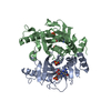
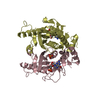
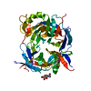
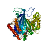
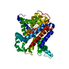
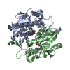
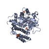
- PDB-2xbu: Saccharomyces cerevisiae hypoxanthine-guanine phosphoribosyltrans... -
+ Open data

- Basic information
| Entry | Database: PDB / ID: 2xbu | |||||||||
|---|---|---|---|---|---|---|---|---|---|---|
| Title | Saccharomyces cerevisiae hypoxanthine-guanine phosphoribosyltransferase in complex with GMP (monoclinic crystal form) | |||||||||
 Components Components | HYPOXANTHINE-GUANINE PHOSPHORIBOSYLTRANSFERASE | |||||||||
 Keywords Keywords | TRANSFERASE / GLYCOSYLTRANSFERASE / PURINE SALVAGE / FLIP PEPTIDE-PLANE | |||||||||
| Function / homology |  Function and homology information Function and homology informationXMP salvage / hypoxanthine phosphoribosyltransferase / guanine phosphoribosyltransferase activity / hypoxanthine metabolic process / hypoxanthine phosphoribosyltransferase activity / GMP salvage / IMP salvage / purine ribonucleoside salvage / nucleotide binding / metal ion binding ...XMP salvage / hypoxanthine phosphoribosyltransferase / guanine phosphoribosyltransferase activity / hypoxanthine metabolic process / hypoxanthine phosphoribosyltransferase activity / GMP salvage / IMP salvage / purine ribonucleoside salvage / nucleotide binding / metal ion binding / nucleus / cytoplasm Similarity search - Function | |||||||||
| Biological species |  | |||||||||
| Method |  X-RAY DIFFRACTION / SYNCHROTRON / MOLECULAR REPLACEMENT / Resolution: 1.8 Å X-RAY DIFFRACTION / SYNCHROTRON / MOLECULAR REPLACEMENT / Resolution: 1.8 Å | |||||||||
 Authors Authors | Moynie, L. / Giraud, M.F. / Breton, A. / Boissier, F. / Daignan-Fornier, B. / Dautant, A. | |||||||||
 Citation Citation | Journal: Protein Sci. / Year: 2012 Title: Functional Significance of Four Successive Glycine Residues in the Pyrophosphate Binding Loop of Fungal 6-Oxopurine Phosphoribosyltransferases. Authors: Moynie, L. / Giraud, M.F. / Breton, A. / Boissier, F. / Daignan-Fornier, B. / Dautant, A. #1: Journal: Genetics / Year: 2008 Title: Lethal Accumulation of Guanylic Nucleotides in Saccharomyces Cerevisiae Hpt1-Deregulated Mutants. Authors: Breton, A. / Pinson, B. / Coulpier, F. / Giraud, M. / Dautant, A. / Daignan-Fornier, B. | |||||||||
| History |
|
- Structure visualization
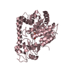
| Structure viewer | Molecule: MolmilJmol/JSmol |
|---|
- Downloads & links
-Download
| PDBx/mmCIF format | 2xbu.cif.gz | 185.6 KB | Display | PDBx/mmCIF format |
|---|---|---|---|---|
| PDB format | pdb2xbu.ent.gz | 147.3 KB | Display | PDB format |
| PDBx/mmJSON format | 2xbu.json.gz | Tree view | PDBx/mmJSON format | |
| Others |  Other downloads Other downloads |
-Validation report
| Arichive directory | https://data.pdbj.org/pub/pdb/validation_reports/xb/2xbuftp://data.pdbj.org/pub/pdb/validation_reports/xb/2xbu | HTTPS FTP |
|---|
-Related structure data
| Related structure data |  2jkySC  2jkzC S: Starting model for refinement C: citing same article ( |
|---|---|
| Similar structure data |


-Links
 PDBj
PDBj

- Assembly
| Deposited unit | 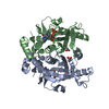
| ||||||||
|---|---|---|---|---|---|---|---|---|---|
| 1 |
| ||||||||
| Unit cell |
| ||||||||
| Noncrystallographic symmetry (NCS) | NCS oper: (Code: given Matrix: (0.9848, 0.01737, 0.173), Vector: |
-Components
| #1: Protein | Mass: 25223.715 Da / Num. of mol.: 2 Source method: isolated from a genetically manipulated source Source: (gene. exp.) Strain: Y1846 / Plasmid: PET-21 / Production host:  References: UniProt: Q04178, hypoxanthine phosphoribosyltransferase #2: Chemical |   Mass: 59.044 Da / Num. of mol.: 2 / Source method: obtained synthetically / Formula: C2H3O2 Mass: 59.044 Da / Num. of mol.: 2 / Source method: obtained synthetically / Formula: C2H3O2#3: Chemical |   Mass: 363.221 Da / Num. of mol.: 2 / Source method: obtained synthetically / Formula: C10H14N5O8P Mass: 363.221 Da / Num. of mol.: 2 / Source method: obtained synthetically / Formula: C10H14N5O8P#4: Chemical | ChemComp-MG / |   Mass: 24.305 Da / Num. of mol.: 1 / Source method: obtained synthetically / Formula: Mg Mass: 24.305 Da / Num. of mol.: 1 / Source method: obtained synthetically / Formula: Mg#5: Water | ChemComp-HOH / |  Mass: 18.015 Da / Num. of mol.: 475 / Source method: isolated from a natural source / Formula: H2O Mass: 18.015 Da / Num. of mol.: 475 / Source method: isolated from a natural source / Formula: H2OHas protein modification | N | |
|---|
-Experimental details
-Experiment
| Experiment | Method: X-RAY DIFFRACTION / Number of used crystals: 1 |
|---|
- Sample preparation
| Crystal | Density Matthews: 1.35 Å3/Da / Density % sol: 45.1 % / Description: NONE |
|---|---|
| Crystal grow | Temperature: 293 K / Method: vapor diffusion, sitting drop / pH: 5.6 Details: 0.2 M AMMONIUM ACETATE, 30% PEG 4000, 0.1 M TRI-SODIUM CITRATE, VAPOR DIFFUSION, SITTING DROP, TEMPERATURE 293K, pH 5.6 |
-Data collection
| Diffraction | Mean temperature: 100 K |
|---|---|
| Diffraction source | Source: SYNCHROTRON / Site: ESRF  / Beamline: ID29 / Wavelength: 0.934 / Beamline: ID29 / Wavelength: 0.934 |
| Detector | Type: ADSC CCD / Detector: CCD / Date: Feb 27, 2008 / Details: MIRRORS |
| Radiation | Monochromator: SI(111) SINGLE CRYSTAL / Protocol: SINGLE WAVELENGTH / Monochromatic (M) / Laue (L): M / Scattering type: x-ray |
| Radiation wavelength | Wavelength: 0.934 Å / Relative weight: 1 |
| Reflection | Resolution: 1.8→28.21 Å / Num. obs: 39543 / % possible obs: 96.6 % / Observed criterion σ(I): 0 / Redundancy: 4.3 % / Biso Wilson estimate: 19.57 Å2 / Rmerge(I) obs: 0.06 / Net I/σ(I): 15.3 |
| Reflection shell | Resolution: 1.8→1.9 Å / Redundancy: 3.6 % / Rmerge(I) obs: 0.37 / Mean I/σ(I) obs: 3 / % possible all: 98.4 |
- Processing
| Software |
| |||||||||||||||||||||||||||||||||||||||||||||||||||||||||||||||||||||||||||||||||||||||||||||||||||||||||
|---|---|---|---|---|---|---|---|---|---|---|---|---|---|---|---|---|---|---|---|---|---|---|---|---|---|---|---|---|---|---|---|---|---|---|---|---|---|---|---|---|---|---|---|---|---|---|---|---|---|---|---|---|---|---|---|---|---|---|---|---|---|---|---|---|---|---|---|---|---|---|---|---|---|---|---|---|---|---|---|---|---|---|---|---|---|---|---|---|---|---|---|---|---|---|---|---|---|---|---|---|---|---|---|---|---|---|
| Refinement | Method to determine structure: MOLECULAR REPLACEMENT Starting model: PDB ENTRY 2JKY Resolution: 1.8→28.214 Å / SU ML: 0.22 / σ(F): 1.36 / Phase error: 19.97 / Stereochemistry target values: ML
| |||||||||||||||||||||||||||||||||||||||||||||||||||||||||||||||||||||||||||||||||||||||||||||||||||||||||
| Solvent computation | Shrinkage radii: 0.9 Å / VDW probe radii: 1.11 Å / Solvent model: FLAT BULK SOLVENT MODEL / Bsol: 63.25 Å2 / ksol: 0.393 e/Å3 | |||||||||||||||||||||||||||||||||||||||||||||||||||||||||||||||||||||||||||||||||||||||||||||||||||||||||
| Displacement parameters | Biso mean: 25.6 Å2
| |||||||||||||||||||||||||||||||||||||||||||||||||||||||||||||||||||||||||||||||||||||||||||||||||||||||||
| Refinement step | Cycle: LAST / Resolution: 1.8→28.214 Å
| |||||||||||||||||||||||||||||||||||||||||||||||||||||||||||||||||||||||||||||||||||||||||||||||||||||||||
| Refine LS restraints |
| |||||||||||||||||||||||||||||||||||||||||||||||||||||||||||||||||||||||||||||||||||||||||||||||||||||||||
| LS refinement shell |
| |||||||||||||||||||||||||||||||||||||||||||||||||||||||||||||||||||||||||||||||||||||||||||||||||||||||||
| Refinement TLS params. | Method: refined / Refine-ID: X-RAY DIFFRACTION
| |||||||||||||||||||||||||||||||||||||||||||||||||||||||||||||||||||||||||||||||||||||||||||||||||||||||||
| Refinement TLS group |
|
