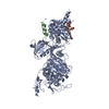
 Movie
Movie Controller


+ Open data

- Basic information
| Entry | Database: PDB / ID: 3j25 | ||||||
|---|---|---|---|---|---|---|---|
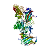
| Title | Structural basis for TetM-mediated tetracycline resistance | ||||||
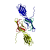
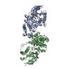
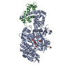
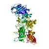
 Components Components | Tetracycline resistance protein tetM | ||||||
 Keywords Keywords | TRANSLATION / antibiotic resistance | ||||||
| Function / homology |  Function and homology information Function and homology informationribosome disassembly / translation / response to antibiotic / GTPase activity / GTP binding Similarity search - Function | ||||||
| Biological species |   Enterococcus faecalis (bacteria) Enterococcus faecalis (bacteria) | ||||||
| Method | ELECTRON MICROSCOPY / single particle reconstruction / cryo EM / Resolution: 7.2 Å | ||||||
 Authors Authors | Doenhoefer, A. / Franckenberg, S. / Wickles, S. / Berninghausen, O. / Beckmann, R. / Wilson, D.N. | ||||||
 Citation Citation | Journal: Proc Natl Acad Sci U S A / Year: 2012 Title: Structural basis for TetM-mediated tetracycline resistance. Authors: Alexandra Dönhöfer / Sibylle Franckenberg / Stephan Wickles / Otto Berninghausen / Roland Beckmann / Daniel N Wilson /  Abstract: Ribosome protection proteins (RPPs) confer tetracycline resistance by binding to the ribosome and chasing the drug from its binding site. The current model for the mechanism of action of RPPs ...Ribosome protection proteins (RPPs) confer tetracycline resistance by binding to the ribosome and chasing the drug from its binding site. The current model for the mechanism of action of RPPs proposes that drug release is indirect and achieved via conformational changes within the drug-binding site induced upon binding of the RPP to the ribosome. Here we report a cryo-EM structure of the RPP TetM in complex with the 70S ribosome at 7.2-Å resolution. The structure reveals the contacts of TetM with the ribosome, including interaction between the conserved and functionally critical C-terminal extension of TetM and the decoding center of the small subunit. Moreover, we observe direct interaction between domain IV of TetM and the tetracycline binding site and identify residues critical for conferring tetracycline resistance. A model is presented whereby TetM directly dislodges tetracycline to confer resistance. | ||||||
| History |
|
- Structure visualization
| Movie |
Movie viewer |
|---|---|
| Structure viewer | Molecule: MolmilJmol/JSmol |
- Downloads & links
-Download
| PDBx/mmCIF format | 3j25.cif.gz | 105.2 KB | Display | PDBx/mmCIF format |
|---|---|---|---|---|
| PDB format | pdb3j25.ent.gz | 63.9 KB | Display | PDB format |
| PDBx/mmJSON format | 3j25.json.gz | Tree view | PDBx/mmJSON format | |
| Others |  Other downloads Other downloads |
-Validation report
| Arichive directory | https://data.pdbj.org/pub/pdb/validation_reports/j2/3j25ftp://data.pdbj.org/pub/pdb/validation_reports/j2/3j25 | HTTPS FTP |
|---|
-Related structure data
| Related structure data |  2183MC M: map data used to model this data C: citing same article ( |
|---|---|
| Similar structure data |


-Links
 PDBj
PDBj


- Assembly
| Deposited unit | 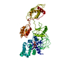
|
|---|---|
| 1 |
|
-Components
| #1: Protein | Mass: 72439.766 Da / Num. of mol.: 1 Source method: isolated from a genetically manipulated source Source: (gene. exp.) Enterococcus faecalis (bacteria) / Gene: tetM, transposon TnFO1 / Plasmid: pET-46 EK/LIC / Production host: |
|---|---|
| #2: Chemical | ChemComp-GCP /   Mass: 521.208 Da / Num. of mol.: 1 / Source method: obtained synthetically / Formula: C11H18N5O13P3 / Comment: GMP-PCP, energy-carrying molecule analogue*YM Mass: 521.208 Da / Num. of mol.: 1 / Source method: obtained synthetically / Formula: C11H18N5O13P3 / Comment: GMP-PCP, energy-carrying molecule analogue*YM |
-Experimental details
-Experiment
| Experiment | Method: ELECTRON MICROSCOPY |
|---|---|
| EM experiment | Aggregation state: PARTICLE / 3D reconstruction method: single particle reconstruction |
- Sample preparation
| Component |
| ||||||||||||
|---|---|---|---|---|---|---|---|---|---|---|---|---|---|
| Buffer solution | Name: 20mM HEPES-KOH, pH 7.8, 30 mM NH4Cl, 10 mM MgCl2 / pH: 7.8 / Details: 20mM HEPES-KOH, pH 7.8, 30 mM NH4Cl, 10 mM MgCl2 | ||||||||||||
| Specimen | Embedding applied: NO / Shadowing applied: NO / Staining applied: NO / Vitrification applied: YES | ||||||||||||
| Vitrification | Instrument: FEI VITROBOT MARK IV / Cryogen name: ETHANE / Chamber temperature: 277 K / Details: liquid ethane / Vitrobot Mark IV |
- Electron microscopy imaging
| Experimental equipment |  Model: Titan Krios / Image courtesy: FEI Company |
|---|---|
| Microscopy | Model: FEI TITAN KRIOS / Date: Jul 1, 2011 |
| Electron gun | Electron source:  FIELD EMISSION GUN / Accelerating voltage: 200 kV / Illumination mode: FLOOD BEAM FIELD EMISSION GUN / Accelerating voltage: 200 kV / Illumination mode: FLOOD BEAM |
| Electron lens | Mode: BRIGHT FIELD / Nominal magnification: 75000 X / Nominal defocus max: -3500 nm / Nominal defocus min: -1000 nm |
| Image recording | Electron dose: 20 e/Å2 / Film or detector model: TVIPS TEMCAM-F416 (4k x 4k) |
| Radiation | Protocol: SINGLE WAVELENGTH / Monochromatic (M) / Laue (L): M / Scattering type: x-ray |
| Radiation wavelength | Relative weight: 1 |
- Processing
| EM software |
| |||||||||||||||
|---|---|---|---|---|---|---|---|---|---|---|---|---|---|---|---|---|
| CTF correction | Details: The volumes were CTF-corrected in defocus groups | |||||||||||||||
| Symmetry | Point symmetry: C1 (asymmetric) | |||||||||||||||
| 3D reconstruction | Method: back-projection interpolated in Fourier space / Resolution: 7.2 Å / Num. of particles: 52701 / Nominal pixel size: 1.04 Å / Actual pixel size: 1.04 Å Details: a modified version of SPIDER program was used for the reconstruction Symmetry type: POINT | |||||||||||||||
| Atomic model building | Protocol: FLEXIBLE FIT / Space: REAL / Target criteria: Cross-correlation coefficient Details: METHOD--Local refinement, Flexible fitting REFINEMENT PROTOCOL--rigid body | |||||||||||||||
| Atomic model building | PDB-ID: 2WRI 2wri Accession code: 2WRI / Source name: PDB / Type: experimental model | |||||||||||||||
| Refinement step | Cycle: LAST
|
