 Movie
Movie Controller

[English] 日本語
 Yorodumi
- PDB-3tew: Crystal Structure of Anthrax Protective Antigen (Membrane Inserti... -
+ Open data

- Basic information
| Entry | Database: PDB / ID: 3tew | ||||||
|---|---|---|---|---|---|---|---|
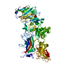
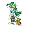
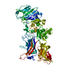
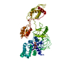
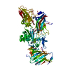
| Title | Crystal Structure of Anthrax Protective Antigen (Membrane Insertion Loop Deleted) to 1.45-A resolution | ||||||
 Components Components | Protective antigen | ||||||
 Keywords Keywords | protein transport / toxin | ||||||
| Function / homology |  Function and homology information Function and homology informationsymbiont-mediated suppression of host MAPK cascade / host cell cytosol / Uptake and function of anthrax toxins / host cell endosome membrane / protein homooligomerization / toxin activity / host cell plasma membrane / extracellular region / metal ion binding / identical protein binding Similarity search - Function | ||||||
| Biological species |  | ||||||
| Method |  X-RAY DIFFRACTION / SYNCHROTRON / MOLECULAR REPLACEMENT / Resolution: 1.45 Å X-RAY DIFFRACTION / SYNCHROTRON / MOLECULAR REPLACEMENT / Resolution: 1.45 Å | ||||||
 Authors Authors | Feld, G.K. / Krantz, B.A. | ||||||
 Citation Citation | Journal: J.Mol.Biol. / Year: 2012 Title: Domain flexibility modulates the heterogeneous assembly mechanism of anthrax toxin protective antigen. Authors: Feld, G.K. / Kintzer, A.F. / Tang, I.I. / Thoren, K.L. / Krantz, B.A. | ||||||
| History |
|
- Structure visualization
| Structure viewer | Molecule: MolmilJmol/JSmol |
|---|
- Downloads & links
-Download
| PDBx/mmCIF format | 3tew.cif.gz | 307.5 KB | Display | PDBx/mmCIF format |
|---|---|---|---|---|
| PDB format | pdb3tew.ent.gz | 247.9 KB | Display | PDB format |
| PDBx/mmJSON format | 3tew.json.gz | Tree view | PDBx/mmJSON format | |
| Others |  Other downloads Other downloads |
-Validation report
| Arichive directory | https://data.pdbj.org/pub/pdb/validation_reports/te/3tewftp://data.pdbj.org/pub/pdb/validation_reports/te/3tew | HTTPS FTP |
|---|
-Related structure data
| Related structure data |  3texC  3teyC  3tezC  1accS C: citing same article ( S: Starting model for refinement |
|---|---|
| Similar structure data |


-Links
 PDBj
PDBj


- Assembly
| Deposited unit | 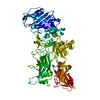
| ||||||||
|---|---|---|---|---|---|---|---|---|---|
| 1 |
| ||||||||
| Unit cell |
|
-Components
| #1: Protein | Mass: 80733.641 Da / Num. of mol.: 1 / Mutation: V303P,H304G Source method: isolated from a genetically manipulated source Source: (gene. exp.) | ||||||
|---|---|---|---|---|---|---|---|
| #2: Chemical |   Mass: 40.078 Da / Num. of mol.: 2 / Source method: obtained synthetically / Formula: Ca Mass: 40.078 Da / Num. of mol.: 2 / Source method: obtained synthetically / Formula: Ca#3: Chemical |   Mass: 76.094 Da / Num. of mol.: 3 / Source method: obtained synthetically / Formula: C3H8O2 Mass: 76.094 Da / Num. of mol.: 3 / Source method: obtained synthetically / Formula: C3H8O2#4: Water | ChemComp-HOH / |  Mass: 18.015 Da / Num. of mol.: 348 / Source method: isolated from a natural source / Formula: H2O Mass: 18.015 Da / Num. of mol.: 348 / Source method: isolated from a natural source / Formula: H2OSequence details | THE CLOSEST DATABASE REFERENCE TO THE CRYSTALLIZED SEQUENCE CORRESPONDS TO UNP ENTRY P13423, WHICH ...THE CLOSEST DATABASE REFERENCE TO THE CRYSTALLIZ | |
-Experimental details
-Experiment
| Experiment | Method: X-RAY DIFFRACTION / Number of used crystals: 1 |
|---|
- Sample preparation
| Crystal | Density Matthews: 2.44 Å3/Da / Density % sol: 49.52 % |
|---|---|
| Crystal grow | Temperature: 292 K / Method: vapor diffusion, hanging drop / pH: 8.5 Details: 20% PEG-ME 2000, 0.1M Tris-Cl, 0.2M Trimethylamine-N-oxide, pH 8.5, VAPOR DIFFUSION, HANGING DROP, temperature 292K |
-Data collection
| Diffraction | Mean temperature: 100 K |
|---|---|
| Diffraction source | Source: SYNCHROTRON / Site: ALS  / Beamline: 8.3.1 / Wavelength: 1.1159 Å / Beamline: 8.3.1 / Wavelength: 1.1159 Å |
| Detector | Type: ADSC QUANTUM 315r / Detector: CCD / Date: Oct 7, 2009 |
| Radiation | Monochromator: Synchrotron / Protocol: SINGLE WAVELENGTH / Monochromatic (M) / Laue (L): M / Scattering type: x-ray |
| Radiation wavelength | Wavelength: 1.1159 Å / Relative weight: 1 |
| Reflection | Resolution: 1.45→23 Å / Num. all: 126263 / Num. obs: 126263 / % possible obs: 90.3 % / Observed criterion σ(F): 1.9 / Observed criterion σ(I): 1.9 / Redundancy: 6.5 % / Biso Wilson estimate: 27 Å2 / Rmerge(I) obs: 0.096 / Rsym value: 0.096 / Net I/σ(I): 13.9 |
| Reflection shell | Resolution: 1.45→1.49 Å / Redundancy: 3.4 % / Rmerge(I) obs: 0.748 / Mean I/σ(I) obs: 1.9 / Num. unique all: 7983 / Rsym value: 0.748 / % possible all: 81 |
- Processing
| Software |
| |||||||||||||||||||||||||||||||||||||||||||||||||||||||||||||||||||||||||||||||||||||||||||||||||||||||||
|---|---|---|---|---|---|---|---|---|---|---|---|---|---|---|---|---|---|---|---|---|---|---|---|---|---|---|---|---|---|---|---|---|---|---|---|---|---|---|---|---|---|---|---|---|---|---|---|---|---|---|---|---|---|---|---|---|---|---|---|---|---|---|---|---|---|---|---|---|---|---|---|---|---|---|---|---|---|---|---|---|---|---|---|---|---|---|---|---|---|---|---|---|---|---|---|---|---|---|---|---|---|---|---|---|---|---|
| Refinement | Method to determine structure: MOLECULAR REPLACEMENT Starting model: 1ACC Resolution: 1.45→23 Å / SU ML: 0.19 / σ(F): 0.06 / Phase error: 22.88 / Stereochemistry target values: ML
| |||||||||||||||||||||||||||||||||||||||||||||||||||||||||||||||||||||||||||||||||||||||||||||||||||||||||
| Solvent computation | Shrinkage radii: 1.37 Å / VDW probe radii: 1.4 Å / Solvent model: FLAT BULK SOLVENT MODEL / Bsol: 40 Å2 / ksol: 0.323 e/Å3 | |||||||||||||||||||||||||||||||||||||||||||||||||||||||||||||||||||||||||||||||||||||||||||||||||||||||||
| Displacement parameters |
| |||||||||||||||||||||||||||||||||||||||||||||||||||||||||||||||||||||||||||||||||||||||||||||||||||||||||
| Refinement step | Cycle: LAST / Resolution: 1.45→23 Å
| |||||||||||||||||||||||||||||||||||||||||||||||||||||||||||||||||||||||||||||||||||||||||||||||||||||||||
| Refine LS restraints |
| |||||||||||||||||||||||||||||||||||||||||||||||||||||||||||||||||||||||||||||||||||||||||||||||||||||||||
| LS refinement shell |
|
