 Movie
Movie Controller

[English] 日本語
 Yorodumi
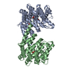
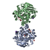
- PDB-6y74: X-ray crystal structure of human carbonic anhydrase IX catalytic ... -
+ Open data

- Basic information
| Entry | Database: PDB / ID: 6y74 | ||||||
|---|---|---|---|---|---|---|---|
| Title | X-ray crystal structure of human carbonic anhydrase IX catalytic domain. | ||||||
 Components Components | Carbonic anhydrase 9 | ||||||
 Keywords Keywords | LYASE / carbonic anhydrase / proton transfer | ||||||
| Function / homology |  Function and homology information Function and homology informationRegulation of gene expression by Hypoxia-inducible Factor / response to testosterone / microvillus membrane / secretion / Reversible hydration of carbon dioxide / molecular function activator activity / morphogenesis of an epithelium / carbonic anhydrase / carbonate dehydratase activity / basolateral plasma membrane ...Regulation of gene expression by Hypoxia-inducible Factor / response to testosterone / microvillus membrane / secretion / Reversible hydration of carbon dioxide / molecular function activator activity / morphogenesis of an epithelium / carbonic anhydrase / carbonate dehydratase activity / basolateral plasma membrane / response to hypoxia / response to xenobiotic stimulus / nucleolus / zinc ion binding / membrane / plasma membrane Similarity search - Function | ||||||
| Biological species |  Homo sapiens (human) Homo sapiens (human) | ||||||
| Method |  X-RAY DIFFRACTION / SYNCHROTRON / MOLECULAR REPLACEMENT / Resolution: 1.53 Å X-RAY DIFFRACTION / SYNCHROTRON / MOLECULAR REPLACEMENT / Resolution: 1.53 Å | ||||||
 Authors Authors | Fisher, S.Z. / Koruza, K. | ||||||
 Citation Citation | Journal: Int J Mol Sci / Year: 2020 Title: Biophysical Characterization of Cancer-Related Carbonic Anhydrase IX Authors: Koruza, K. / Murray, A.B. / Mahon, B.P. / Hopkins, J.B. / Knecht, W. / McKenna, R. / Fisher, S.Z. | ||||||
| History |
|
- Structure visualization
| Structure viewer | Molecule: MolmilJmol/JSmol |
|---|
- Downloads & links
-Download
| PDBx/mmCIF format | 6y74.cif.gz | 134.6 KB | Display | PDBx/mmCIF format |
|---|---|---|---|---|
| PDB format | pdb6y74.ent.gz | 102.5 KB | Display | PDB format |
| PDBx/mmJSON format | 6y74.json.gz | Tree view | PDBx/mmJSON format | |
| Others |  Other downloads Other downloads |
-Validation report
| Arichive directory | https://data.pdbj.org/pub/pdb/validation_reports/y7/6y74ftp://data.pdbj.org/pub/pdb/validation_reports/y7/6y74 | HTTPS FTP |
|---|
-Related structure data
| Related structure data | 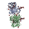 3iaiS S: Starting model for refinement |
|---|---|
| Similar structure data |


-Links
 PDBj
PDBj


- Assembly
| Deposited unit | 
| ||||||||
|---|---|---|---|---|---|---|---|---|---|
| 1 |
| ||||||||
| Unit cell |
|
-Components
-Protein , 1 types, 2 molecules AB
| #1: Protein | Mass: 28261.844 Da / Num. of mol.: 2 Source method: isolated from a genetically manipulated source Source: (gene. exp.) Homo sapiens (human) / Gene: CA9, G250, MN / Plasmid: Bac-to-BacProduction host:  Spodoptera aff. frugiperda 1 BOLD-2017 (butterflies/moths) Spodoptera aff. frugiperda 1 BOLD-2017 (butterflies/moths)References: UniProt: Q16790, carbonic anhydrase |
|---|
-Sugars , 2 types, 2 molecules
| #2: Polysaccharide | beta-D-mannopyranose-(1-4)-2-acetamido-2-deoxy-beta-D-glucopyranose-(1-4)-2-acetamido-2-deoxy-beta- ...beta-D-mannopyranose-(1-4)-2-acetamido-2-deoxy-beta-D-glucopyranose-(1-4)-2-acetamido-2-deoxy-beta-D-glucopyranose Source method: isolated from a genetically manipulated source |
|---|---|
| #3: Polysaccharide | 2-acetamido-2-deoxy-beta-D-glucopyranose-(1-4)-2-acetamido-2-deoxy-beta-D-glucopyranose Source method: isolated from a genetically manipulated source |
-Non-polymers , 4 types, 584 molecules 


| #4: Chemical |  Mass: 65.409 Da / Num. of mol.: 2 / Source method: obtained synthetically / Formula: Zn / Feature type: SUBJECT OF INVESTIGATION Mass: 65.409 Da / Num. of mol.: 2 / Source method: obtained synthetically / Formula: Zn / Feature type: SUBJECT OF INVESTIGATION#5: Chemical |  Mass: 35.453 Da / Num. of mol.: 3 / Source method: obtained synthetically / Formula: Cl Mass: 35.453 Da / Num. of mol.: 3 / Source method: obtained synthetically / Formula: Cl#6: Chemical |  Mass: 59.044 Da / Num. of mol.: 2 / Source method: obtained synthetically / Formula: C2H3O2 Mass: 59.044 Da / Num. of mol.: 2 / Source method: obtained synthetically / Formula: C2H3O2#7: Water | ChemComp-HOH / | Mass: 18.015 Da / Num. of mol.: 577 / Source method: isolated from a natural source / Formula: H2O |
|---|
-Details
| Has ligand of interest | Y |
|---|---|
| Has protein modification | Y |
-Experimental details
-Experiment
| Experiment | Method: X-RAY DIFFRACTION / Number of used crystals: 1 |
|---|
- Sample preparation
| Crystal | Density Matthews: 2.3 Å3/Da / Density % sol: 46.64 % |
|---|---|
| Crystal grow | Temperature: 293 K / Method: vapor diffusion, sitting drop / pH: 4.3 Details: 17% PEG 6000, 1.4 M Sodium citrate pH 4.3, 1 M LiCl |
-Data collection
| Diffraction | Mean temperature: 100 K / Serial crystal experiment: N |
|---|---|
| Diffraction source | Source: SYNCHROTRON / Site: MAX IV  / Beamline: BioMAX / Wavelength: 0.979 Å / Beamline: BioMAX / Wavelength: 0.979 Å |
| Detector | Type: DECTRIS EIGER X 16M / Detector: PIXEL / Date: May 8, 2018 |
| Radiation | Protocol: SINGLE WAVELENGTH / Monochromatic (M) / Laue (L): M / Scattering type: x-ray |
| Radiation wavelength | Wavelength: 0.979 Å / Relative weight: 1 |
| Reflection | Resolution: 1.53→37.13 Å / Num. obs: 75610 / % possible obs: 98.3 % / Redundancy: 6.9 % / Biso Wilson estimate: 15.6 Å2 / Rmerge(I) obs: 0.11 / Net I/σ(I): 10.6 |
| Reflection shell | Resolution: 1.53→1.56 Å / Rmerge(I) obs: 0.815 / Mean I/σ(I) obs: 2.2 / Num. unique obs: 3288 |
- Processing
| Software |
| |||||||||||||||||||||||||||||||||||||||||||||||||||||||||||||||||||||||||||||||||||||||||||||||||||||||||||||||||||||||||||||||||||||||||||||||||||||||||||||||||||||||||||||||||||||||||||||
|---|---|---|---|---|---|---|---|---|---|---|---|---|---|---|---|---|---|---|---|---|---|---|---|---|---|---|---|---|---|---|---|---|---|---|---|---|---|---|---|---|---|---|---|---|---|---|---|---|---|---|---|---|---|---|---|---|---|---|---|---|---|---|---|---|---|---|---|---|---|---|---|---|---|---|---|---|---|---|---|---|---|---|---|---|---|---|---|---|---|---|---|---|---|---|---|---|---|---|---|---|---|---|---|---|---|---|---|---|---|---|---|---|---|---|---|---|---|---|---|---|---|---|---|---|---|---|---|---|---|---|---|---|---|---|---|---|---|---|---|---|---|---|---|---|---|---|---|---|---|---|---|---|---|---|---|---|---|---|---|---|---|---|---|---|---|---|---|---|---|---|---|---|---|---|---|---|---|---|---|---|---|---|---|---|---|---|---|---|---|---|
| Refinement | Method to determine structure: MOLECULAR REPLACEMENT Starting model: 3iai Resolution: 1.53→37.13 Å / SU ML: 0.16 / Cross valid method: THROUGHOUT / σ(F): 1.36 / Phase error: 21.22
| |||||||||||||||||||||||||||||||||||||||||||||||||||||||||||||||||||||||||||||||||||||||||||||||||||||||||||||||||||||||||||||||||||||||||||||||||||||||||||||||||||||||||||||||||||||||||||||
| Solvent computation | Shrinkage radii: 0.9 Å / VDW probe radii: 1.11 Å | |||||||||||||||||||||||||||||||||||||||||||||||||||||||||||||||||||||||||||||||||||||||||||||||||||||||||||||||||||||||||||||||||||||||||||||||||||||||||||||||||||||||||||||||||||||||||||||
| Displacement parameters | Biso max: 112.55 Å2 / Biso mean: 21.5263 Å2 / Biso min: 7.9 Å2 | |||||||||||||||||||||||||||||||||||||||||||||||||||||||||||||||||||||||||||||||||||||||||||||||||||||||||||||||||||||||||||||||||||||||||||||||||||||||||||||||||||||||||||||||||||||||||||||
| Refinement step | Cycle: final / Resolution: 1.53→37.13 Å
| |||||||||||||||||||||||||||||||||||||||||||||||||||||||||||||||||||||||||||||||||||||||||||||||||||||||||||||||||||||||||||||||||||||||||||||||||||||||||||||||||||||||||||||||||||||||||||||
| LS refinement shell | Refine-ID: X-RAY DIFFRACTION / Rfactor Rfree error: 0 / Total num. of bins used: 26
|
