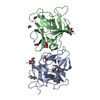
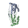
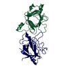
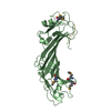
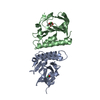
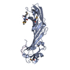
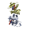
[English] 日本語
Yorodumi
- PDB-6xwg: Crystal Structure of the Human RXR/RAR DNA-Binding Domain Heterod... -

+
Open data


ID or keywords:

Loading...

-
Basic information

Entry
Database: PDB / ID: 6xwg
TitleCrystal Structure of the Human RXR/RAR DNA-Binding Domain Heterodimer Bound to the Human RARb2 DR5 Response Element
Components
  • (RARb2 DR5 Response Element, ...) x 2
  • (Retinoic acid receptor ...) x 2
KeywordsTRANSCRIPTION / Protein-DNA complex / Nuclear Receptor
Function / homology
Function and homology information


Sertoli cell fate commitment / positive regulation of binding / trachea cartilage development / ventricular cardiac muscle cell differentiation / chondroblast differentiation / embryonic camera-type eye development / protein kinase B binding / negative regulation of granulocyte differentiation / growth plate cartilage development / glandular epithelial cell development ...Sertoli cell fate commitment / positive regulation of binding / trachea cartilage development / ventricular cardiac muscle cell differentiation / chondroblast differentiation / embryonic camera-type eye development / protein kinase B binding / negative regulation of granulocyte differentiation / growth plate cartilage development / glandular epithelial cell development / positive regulation of T-helper 2 cell differentiation / prostate gland development / positive regulation of transporter activity / negative regulation of cartilage development / retinoic acid-responsive element binding / NR1H2 & NR1H3 regulate gene expression linked to triglyceride lipolysis in adipose / NR1H2 & NR1H3 regulate gene expression to limit cholesterol uptake / NR1H2 & NR1H3 regulate gene expression linked to gluconeogenesis / regulation of hematopoietic progenitor cell differentiation / positive regulation of thyroid hormone receptor signaling pathway / positive regulation of interleukin-13 production / positive regulation of interleukin-5 production / NR1H2 & NR1H3 regulate gene expression linked to lipogenesis / Carnitine shuttle / outflow tract septum morphogenesis / retinoic acid binding / TGFBR3 expression / positive regulation of vitamin D receptor signaling pathway / nuclear vitamin D receptor binding / response to vitamin A / limb development / apoptotic cell clearance / regulation of myelination / Signaling by Retinoic Acid / ureteric bud development / DNA binding domain binding / DNA-binding transcription repressor activity / NR1H2 & NR1H3 regulate gene expression to control bile acid homeostasis / protein kinase A binding / nuclear steroid receptor activity / heterocyclic compound binding / LBD domain binding / positive regulation of interleukin-4 production / alpha-actinin binding / face development / germ cell development / negative regulation of type II interferon production / Synthesis of bile acids and bile salts / negative regulation of tumor necrosis factor production / cellular response to estrogen stimulus / positive regulation of cholesterol efflux / Synthesis of bile acids and bile salts via 27-hydroxycholesterol / Endogenous sterols / Synthesis of bile acids and bile salts via 7alpha-hydroxycholesterol / positive regulation of bone mineralization / response to retinoic acid / Transcriptional regulation of brown and beige adipocyte differentiation by EBF2 / retinoic acid receptor signaling pathway / Recycling of bile acids and salts / NR1H3 & NR1H2 regulate gene expression linked to cholesterol transport and efflux / cellular response to retinoic acid / positive regulation of cell cycle / hormone-mediated signaling pathway / : / Regulation of lipid metabolism by PPARalpha / peroxisome proliferator activated receptor signaling pathway / response to cytokine / positive regulation of neuron differentiation / BMAL1:CLOCK,NPAS2 activates circadian expression / mRNA regulatory element binding translation repressor activity / negative regulation of miRNA transcription / peptide binding / Activation of gene expression by SREBF (SREBP) / liver development / hippocampus development / transcription coregulator binding / RNA polymerase II transcription regulatory region sequence-specific DNA binding / female pregnancy / neural tube closure / SUMOylation of intracellular receptors / mRNA transcription by RNA polymerase II / Heme signaling / Transcriptional activation of mitochondrial biogenesis / transcription coactivator binding / PPARA activates gene expression / Cytoprotection by HMOX1 / multicellular organism growth / regulation of synaptic plasticity / Nuclear Receptor transcription pathway / chromatin DNA binding / Transcriptional regulation of white adipocyte differentiation / mRNA 5'-UTR binding / Activation of anterior HOX genes in hindbrain development during early embryogenesis / histone deacetylase binding / RNA polymerase II transcription regulator complex / Transcriptional regulation of granulopoiesis / nuclear receptor activity / sequence-specific double-stranded DNA binding / : / nervous system development
Similarity search - Function
: / : / Retinoic acid receptor / Nuclear/hormone receptor activator site AF-1 / Nuclear/hormone receptor activator site AF-1 / Retinoid X receptor/HNF4 / : / Nuclear hormone receptor / Nuclear hormones receptors DNA-binding region signature. / Zinc finger, nuclear hormone receptor-type ...: / : / Retinoic acid receptor / Nuclear/hormone receptor activator site AF-1 / Nuclear/hormone receptor activator site AF-1 / Retinoid X receptor/HNF4 / : / Nuclear hormone receptor / Nuclear hormones receptors DNA-binding region signature. / Zinc finger, nuclear hormone receptor-type / Double treble clef zinc finger, C4 type / Nuclear hormone receptors DNA-binding domain profile. / c4 zinc finger in nuclear hormone receptors / Nuclear hormone receptor, ligand-binding domain / Nuclear hormone receptor-like domain superfamily / Ligand-binding domain of nuclear hormone receptor / Nuclear receptor (NR) ligand-binding (LBD) domain profile. / Ligand binding domain of hormone receptors / Zinc finger, NHR/GATA-type
Similarity search - Domain/homology
DNA / DNA (> 10) / Retinoic acid receptor alpha / Retinoic acid receptor RXR-alpha
Similarity search - Component
Biological speciesHomo sapiens (human)
MethodX-RAY DIFFRACTION / SYNCHROTRON / MOLECULAR REPLACEMENT / molecular replacement / Resolution: 2.4 Å
AuthorsMcEwen, A.G. / Poussin-Courmontagne, P. / Peluso-Iltis, C. / Rochel, N.
Funding support France, 5items
OrganizationGrant numberCountry
French National Research AgencyANR-11-BSV8-023 France
Fondation ARCSFI20121205585 France
French National Research AgencyFRISBI ANR-10-INBS-05 France
French National Research AgencyANR-10-LABX-0030-INRT France
French National Research AgencyANR-10-IDEX-0002-02 France
CitationJournal: Nucleic Acids Res. / Year: 2020
Title: Structural basis for DNA recognition and allosteric control of the retinoic acid receptors RAR-RXR.
Authors: Osz, J. / McEwen, A.G. / Bourguet, M. / Przybilla, F. / Peluso-Iltis, C. / Poussin-Courmontagne, P. / Mely, Y. / Cianferani, S. / Jeffries, C.M. / Svergun, D.I. / Rochel, N.
History
DepositionJan 23, 2020Deposition site: PDBE / Processing site: PDBE
Revision 1.0Sep 9, 2020Provider: repository / Type: Initial release
Revision 1.1Oct 7, 2020Group: Database references / Category: citation / citation_author
Item: _citation.journal_volume / _citation.page_first ..._citation.journal_volume / _citation.page_first / _citation.page_last / _citation.pdbx_database_id_PubMed / _citation.title / _citation_author.identifier_ORCID
Revision 1.2Jan 24, 2024Group: Data collection / Database references / Refinement description
Category: chem_comp_atom / chem_comp_bond ...chem_comp_atom / chem_comp_bond / database_2 / pdbx_initial_refinement_model
Item: _database_2.pdbx_DOI / _database_2.pdbx_database_accession

-
Structure visualization

Structure viewerMolecule:
MolmilJmol/JSmol

Downloads & links

-
Assembly

Deposited unit
A: RARb2 DR5 Response Element, 5'-3' strand
B: RARb2 DR5 Response Element, 3'-5' strand
C: Retinoic acid receptor RXR-alpha
D: Retinoic acid receptor alpha
hetero molecules


Theoretical massNumber of molelcules
Total (without water)34,17110
Polymers33,7824
Non-polymers3896
Water64936
1


  • Idetical with deposited unit
  • defined by author&software
  • Evidence: gel filtration
TypeNameSymmetry operationNumber
identity operation1_555x,y,z1
Buried area5860 Å2
ΔGint-43 kcal/mol
Surface area15760 Å2
MethodPISA
Unit cell
Length a, b, c (Å)56.714, 56.714, 238.364
Angle α, β, γ (deg.)90.000, 90.000, 90.000
Int Tables number96
Space group name H-MP43212

-
Components

-
RARb2 DR5 Response Element, ... , 2 types, 2 molecules AB

#1: DNA chain RARb2 DR5 Response Element, 5'-3' strand


Mass: 6447.185 Da / Num. of mol.: 1 / Source method: obtained synthetically / Source: (synth.) Homo sapiens (human)
#2: DNA chain RARb2 DR5 Response Element, 3'-5' strand


Mass: 6438.171 Da / Num. of mol.: 1 / Source method: obtained synthetically / Source: (synth.) Homo sapiens (human)

-
Retinoic acid receptor ... , 2 types, 2 molecules CD

#3: Protein Retinoic acid receptor RXR-alpha / Nuclear receptor subfamily 2 group B member 1 / Retinoid X receptor alpha


Mass: 10262.926 Da / Num. of mol.: 1
Source method: isolated from a genetically manipulated source
Details: DNA-Binding Domain / Source: (gene. exp.) Homo sapiens (human) / Gene: RXRA, NR2B1 / Production host: Escherichia coli BL21(DE3) (bacteria) / References: UniProt: P19793
#4: Protein Retinoic acid receptor alpha / RAR-alpha / Nuclear receptor subfamily 1 group B member 1


Mass: 10633.478 Da / Num. of mol.: 1
Source method: isolated from a genetically manipulated source
Details: DNA-Binding Domain / Source: (gene. exp.) Homo sapiens (human) / Gene: RARA, NR1B1 / Production host: Escherichia coli BL21(DE3) (bacteria) / References: UniProt: P10276

-
Non-polymers , 4 types, 42 molecules

#5: Chemical
ChemComp-ZN / ZINC ION


Mass: 65.409 Da / Num. of mol.: 4 / Source method: obtained synthetically / Formula: Zn
#6: Chemical ChemComp-GOL / GLYCEROL / GLYCERIN / PROPANE-1,2,3-TRIOL


Mass: 92.094 Da / Num. of mol.: 1 / Source method: obtained synthetically / Formula: C3H8O3
#7: Chemical ChemComp-CL / CHLORIDE ION


Mass: 35.453 Da / Num. of mol.: 1 / Source method: obtained synthetically / Formula: Cl
#8: Water ChemComp-HOH / water


Mass: 18.015 Da / Num. of mol.: 36 / Source method: isolated from a natural source / Formula: H2O

-
Details

Has ligand of interestN

-
Experimental details

-
Experiment

ExperimentMethod: X-RAY DIFFRACTION / Number of used crystals: 1

-
Sample preparation

CrystalDensity Matthews: 2.82 Å3/Da / Density % sol: 59.9 %
Crystal growTemperature: 293 K / Method: vapor diffusion, sitting drop / pH: 6.5 / Details: 25% PEG 3350, 0.2 M Li2SO4, 0.1 M bis-tris pH 6.5

-
Data collection

DiffractionMean temperature: 100 K / Serial crystal experiment: N
Diffraction sourceSource: SYNCHROTRON / Site: ESRF / Beamline: ID23-1 / Wavelength: 1.006 Å
DetectorType: DECTRIS PILATUS 6M / Detector: PIXEL / Date: Nov 21, 2016
RadiationMonochromator: Si(111) / Protocol: SINGLE WAVELENGTH / Monochromatic (M) / Laue (L): M / Scattering type: x-ray
Radiation wavelengthWavelength: 1.006 Å / Relative weight: 1
ReflectionResolution: 2.4→26.51 Å / Num. obs: 16005 / % possible obs: 99.2 % / Redundancy: 9.756 % / Biso Wilson estimate: 81.7 Å2 / CC1/2: 0.999 / Rmerge(I) obs: 0.111 / Rrim(I) all: 0.118 / Χ2: 1.388 / Net I/σ(I): 9.77 / Num. measured all: 156137
Reflection shell

Diffraction-ID: 1

Resolution (Å)Redundancy (%)Rmerge(I) obsMean I/σ(I) obsNum. measured obsNum. possibleNum. unique obsCC1/2Rrim(I) all% possible all
2.4-2.558.8145.9550.2921753252424680.26.29497.8
2.55-2.729.2732.7620.6421847239323560.2982.91798.5
2.72-2.949.8911.421.3121750221821990.6021.599.1
2.94-3.229.5180.5573.4319759208020760.9640.59199.8
3.22-3.610.3850.2169.6419535188618810.9910.22999.7
3.6-4.1510.1980.1416.6917113167916780.9940.14999.9
4.15-5.0810.4870.124.5215406146914690.9960.105100
5.08-7.1610.1650.07729.9911771115911580.9970.08199.9
7.16-26.5110.0040.04937.9172037207200.9990.052100

-
Phasing

PhasingMethod: molecular replacement

-
Processing

Software
NameVersionClassificationNB
XDSdata reduction
XSCALEdata scaling
PHASERphasing
BUSTERrefinement
PDB_EXTRACT3.25data extraction
RefinementMethod to determine structure: MOLECULAR REPLACEMENT
Starting model: 4CN5
Resolution: 2.4→26.51 Å / Cor.coef. Fo:Fc: 0.953 / Cor.coef. Fo:Fc free: 0.957 / Rfactor Rfree error: 0 / SU R Cruickshank DPI: 0.397 / Cross valid method: THROUGHOUT / σ(F): 0 / SU R Blow DPI: 0.486 / SU Rfree Blow DPI: 0.238 / SU Rfree Cruickshank DPI: 0.228
RfactorNum. reflection% reflectionSelection details
Rfree0.207 543 4.45 %RANDOM
Rwork0.191 ---
obs0.192 12203 75.6 %-
Displacement parametersBiso max: 197.39 Å2 / Biso mean: 83.26 Å2 / Biso min: 34.81 Å2
Baniso -1Baniso -2Baniso -3
1--1.7256 Å20 Å20 Å2
2---1.7256 Å20 Å2
3---3.4512 Å2
Refine analyzeLuzzati coordinate error obs: 0 Å
Refinement stepCycle: final / Resolution: 2.4→26.51 Å
ProteinNucleic acidLigandSolventTotal
Num. atoms1295 855 11 36 2197
Biso mean--78.55 59.32 -
Num. residues----201
Refine LS restraints
Refine-IDTypeNumberRestraint functionWeightDev ideal
X-RAY DIFFRACTIONt_dihedral_angle_d715SINUSOIDAL2
X-RAY DIFFRACTIONt_trig_c_planes30HARMONIC2
X-RAY DIFFRACTIONt_gen_planes237HARMONIC5
X-RAY DIFFRACTIONt_it2278HARMONIC20
X-RAY DIFFRACTIONt_nbd4SEMIHARMONIC5
X-RAY DIFFRACTIONt_improper_torsion
X-RAY DIFFRACTIONt_pseud_angle
X-RAY DIFFRACTIONt_chiral_improper_torsion286SEMIHARMONIC5
X-RAY DIFFRACTIONt_sum_occupancies
X-RAY DIFFRACTIONt_utility_distance
X-RAY DIFFRACTIONt_utility_angle
X-RAY DIFFRACTIONt_utility_torsion
X-RAY DIFFRACTIONt_ideal_dist_contact2255SEMIHARMONIC4
X-RAY DIFFRACTIONt_bond_d2278HARMONIC20.01
X-RAY DIFFRACTIONt_angle_deg3224HARMONIC20.98
X-RAY DIFFRACTIONt_omega_torsion2.88
X-RAY DIFFRACTIONt_other_torsion22.96
LS refinement shellResolution: 2.4→2.63 Å / Rfactor Rfree error: 0 / Total num. of bins used: 6
RfactorNum. reflection% reflection
Rfree0.262 37 4.52 %
Rwork0.215 781 -
all0.217 818 -
obs--21.86 %
Refinement TLS params.

Method: refined / Refine-ID: X-RAY DIFFRACTION

IDL112)L122)L132)L222)L232)L332)S11 (Å °)S12 (Å °)S13 (Å °)S21 (Å °)S22 (Å °)S23 (Å °)S31 (Å °)S32 (Å °)S33 (Å °)T112)T122)T132)T222)T232)T332)Origin x (Å)Origin y (Å)Origin z (Å)
16.2718-0.7474-0.53266.2332-1.52626.7539-0.0762-0.228-0.17640.1690.1103-0.0161-0.20850.0196-0.0341-0.04130.152-0.0872-0.02240.0372-0.159114.1964-22.1246-17.5576
28.31550.6765-0.73626.26211.48295.6766-0.11480.1161-0.0425-0.1096-0.10890.54420.1654-0.26180.2237-0.15150.0085-0.0513-0.17460.03360.0924-8.3268-0.3352-31.2499
33.9483-2.14281.17836.0475-2.91044.7873-0.1607-0.54420.14280.5442-0.10220.3519-0.04830.48930.2629-0.18650.152-0.147-0.10430.0847-0.16543.0336-6.8335-11.1681
Refinement TLS group
IDRefine-IDRefine TLS-IDSelection detailsAuth asym-IDAuth seq-ID
1X-RAY DIFFRACTION1{ C|132 - C|302 }C132 - 302
2X-RAY DIFFRACTION2{ D|82 - D|302 }D82 - 302
3X-RAY DIFFRACTION3{ A|1 - A|51 B|1 - B|21 }A1 - 51
4X-RAY DIFFRACTION3{ A|1 - A|51 B|1 - B|21 }B1 - 21

+
About Yorodumi

-
News

-
Feb 9, 2022. New format data for meta-information of EMDB entries

New format data for meta-information of EMDB entries

  • Version 3 of the EMDB header file is now the official format.
  • The previous official version 1.9 will be removed from the archive.

Related info.:EMDB header

External links:wwPDB to switch to version 3 of the EMDB data model

-
Aug 12, 2020. Covid-19 info

Covid-19 info

URL: https://pdbj.org/emnavi/covid19.php

New page: Covid-19 featured information page in EM Navigator.

Related info.:Covid-19 info / Mar 5, 2020. Novel coronavirus structure data

+
Mar 5, 2020. Novel coronavirus structure data

Novel coronavirus structure data

Related info.:Yorodumi Speices / Aug 12, 2020. Covid-19 info

External links:COVID-19 featured content - PDBj / Molecule of the Month (242):Coronavirus Proteases

+
Jan 31, 2019. EMDB accession codes are about to change! (news from PDBe EMDB page)

EMDB accession codes are about to change! (news from PDBe EMDB page)

  • The allocation of 4 digits for EMDB accession codes will soon come to an end. Whilst these codes will remain in use, new EMDB accession codes will include an additional digit and will expand incrementally as the available range of codes is exhausted. The current 4-digit format prefixed with “EMD-” (i.e. EMD-XXXX) will advance to a 5-digit format (i.e. EMD-XXXXX), and so on. It is currently estimated that the 4-digit codes will be depleted around Spring 2019, at which point the 5-digit format will come into force.
  • The EM Navigator/Yorodumi systems omit the EMD- prefix.

Related info.:Q: What is EMD? / ID/Accession-code notation in Yorodumi/EM Navigator

External links:EMDB Accession Codes are Changing Soon! / Contact to PDBj

+
Jul 12, 2017. Major update of PDB

Major update of PDB

  • wwPDB released updated PDB data conforming to the new PDBx/mmCIF dictionary.
  • This is a major update changing the version number from 4 to 5, and with Remediation, in which all the entries are updated.
  • In this update, many items about electron microscopy experimental information are reorganized (e.g. em_software).
  • Now, EM Navigator and Yorodumi are based on the updated data.

External links:wwPDB Remediation / Enriched Model Files Conforming to OneDep Data Standards Now Available in the PDB FTP Archive

-
Yorodumi

Thousand views of thousand structures

  • Yorodumi is a browser for structure data from EMDB, PDB, SASBDB, etc.
  • This page is also the successor to EM Navigator detail page, and also detail information page/front-end page for Omokage search.
  • The word "yorodu" (or yorozu) is an old Japanese word meaning "ten thousand". "mi" (miru) is to see.

Related info.:EMDB / PDB / SASBDB / Comparison of 3 databanks / Yorodumi Search / Aug 31, 2016. New EM Navigator & Yorodumi / Yorodumi Papers / Jmol/JSmol / Function and homology information / Changes in new EM Navigator and Yorodumi

Read more