 Movie
Movie Controller

[English] 日本語
 Yorodumi
- PDB-4cn5: Crystal Structure of the Human Retinoid X Receptor DNA-Binding Do... -
+ Open data

- Basic information
| Entry | Database: PDB / ID: 4cn5 | ||||||
|---|---|---|---|---|---|---|---|
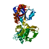
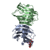
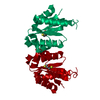
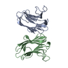
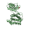
| Title | Crystal Structure of the Human Retinoid X Receptor DNA-Binding Domain Bound to the Human Nr1d1 Response Element | ||||||
 Components Components |
| ||||||
 Keywords Keywords | TRANSCRIPTION/DNA / TRANSCRIPTION-DNA COMPLEX | ||||||
| Function / homology |  Function and homology information Function and homology informationretinoic acid-responsive element binding / NR1H2 & NR1H3 regulate gene expression linked to triglyceride lipolysis in adipose / NR1H2 & NR1H3 regulate gene expression linked to gluconeogenesis / positive regulation of thyroid hormone receptor signaling pathway / NR1H2 & NR1H3 regulate gene expression to limit cholesterol uptake / NR1H2 & NR1H3 regulate gene expression linked to lipogenesis / Carnitine shuttle / retinoic acid binding / positive regulation of vitamin D receptor signaling pathway / TGFBR3 expression ...retinoic acid-responsive element binding / NR1H2 & NR1H3 regulate gene expression linked to triglyceride lipolysis in adipose / NR1H2 & NR1H3 regulate gene expression linked to gluconeogenesis / positive regulation of thyroid hormone receptor signaling pathway / NR1H2 & NR1H3 regulate gene expression to limit cholesterol uptake / NR1H2 & NR1H3 regulate gene expression linked to lipogenesis / Carnitine shuttle / retinoic acid binding / positive regulation of vitamin D receptor signaling pathway / TGFBR3 expression / nuclear vitamin D receptor binding / Signaling by Retinoic Acid / NR1H2 & NR1H3 regulate gene expression to control bile acid homeostasis / DNA binding domain binding / positive regulation of lipid metabolic process / LBD domain binding / positive regulation of lipoprotein transport / nuclear steroid receptor activity / Synthesis of bile acids and bile salts / Synthesis of bile acids and bile salts via 27-hydroxycholesterol / positive regulation of cholesterol efflux / Endogenous sterols / Synthesis of bile acids and bile salts via 7alpha-hydroxycholesterol / monocyte differentiation / response to retinoic acid / cellular response to low-density lipoprotein particle stimulus / positive regulation of bone mineralization / Transcriptional regulation of brown and beige adipocyte differentiation by EBF2 / Recycling of bile acids and salts / retinoic acid receptor signaling pathway / NR1H3 & NR1H2 regulate gene expression linked to cholesterol transport and efflux / Regulation of lipid metabolism by PPARalpha / cell maturation / peptide binding / peroxisome proliferator activated receptor signaling pathway / hormone-mediated signaling pathway / BMAL1:CLOCK,NPAS2 activates circadian expression / RORA,B,C and NR1D1 (REV-ERBA) regulate gene expression / Activation of gene expression by SREBF (SREBP) / Expression of BMAL (ARNTL), CLOCK, and NPAS2 / transcription coregulator binding / SUMOylation of intracellular receptors / RNA polymerase II transcription regulatory region sequence-specific DNA binding / Heme signaling / PPARA activates gene expression / Transcriptional activation of mitochondrial biogenesis / Cytoprotection by HMOX1 / Nuclear Receptor transcription pathway / Transcriptional regulation of white adipocyte differentiation / mRNA transcription by RNA polymerase II / nuclear receptor activity / RNA polymerase II transcription regulator complex / Activation of anterior HOX genes in hindbrain development during early embryogenesis / Transcriptional regulation of granulopoiesis / sequence-specific double-stranded DNA binding / nervous system development / MLL4 and MLL3 complexes regulate expression of PPARG target genes in adipogenesis and hepatic steatosis / double-stranded DNA binding / transcription regulator complex / sequence-specific DNA binding / DNA-binding transcription factor activity, RNA polymerase II-specific / cell differentiation / signaling receptor complex / transcription cis-regulatory region binding / RNA polymerase II cis-regulatory region sequence-specific DNA binding / DNA-binding transcription factor activity / positive regulation of DNA-templated transcription / chromatin / enzyme binding / negative regulation of transcription by RNA polymerase II / positive regulation of transcription by RNA polymerase II / mitochondrion / zinc ion binding / nucleoplasm / identical protein binding / nucleus / cytosol Similarity search - Function | ||||||
| Biological species |  HOMO SAPIENS (human) HOMO SAPIENS (human) | ||||||
| Method |  X-RAY DIFFRACTION / SYNCHROTRON / MOLECULAR REPLACEMENT / Resolution: 2 Å X-RAY DIFFRACTION / SYNCHROTRON / MOLECULAR REPLACEMENT / Resolution: 2 Å | ||||||
 Authors Authors | McEwen, A.G. / Poussin-Courmontagne, P. / Osz, J. / Rochel, N. | ||||||
 Citation Citation | Journal: Sci.Rep. / Year: 2015 Title: Structural Basis of Natural Promoter Recognition by the Retinoid X Nuclear Receptor. Authors: Osz, J. / Mcewen, A.G. / Poussin-Courmontagne, P. / Moutier, E. / Birck, C. / Davidson, I. / Moras, D. / Rochel, N. | ||||||
| History |
|
- Structure visualization
| Structure viewer | Molecule: MolmilJmol/JSmol |
|---|
- Downloads & links
-Download
| PDBx/mmCIF format | 4cn5.cif.gz | 127 KB | Display | PDBx/mmCIF format |
|---|---|---|---|---|
| PDB format | pdb4cn5.ent.gz | 95.9 KB | Display | PDB format |
| PDBx/mmJSON format | 4cn5.json.gz | Tree view | PDBx/mmJSON format | |
| Others |  Other downloads Other downloads |
-Validation report
| Arichive directory | https://data.pdbj.org/pub/pdb/validation_reports/cn/4cn5ftp://data.pdbj.org/pub/pdb/validation_reports/cn/4cn5 | HTTPS FTP |
|---|
-Related structure data
| Related structure data | 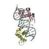 4cn2C  4cn3C  4cn7C  1dszS C: citing same article ( S: Starting model for refinement |
|---|---|
| Similar structure data |


-Links
 PDBj
PDBj


- Assembly
| Deposited unit | 
| ||||||||
|---|---|---|---|---|---|---|---|---|---|
| 1 |
| ||||||||
| Unit cell |
|
-Components
-Protein , 1 types, 2 molecules AB
| #1: Protein | Mass: 10262.926 Da / Num. of mol.: 2 / Fragment: DNA-BINDING DOMAIN, RESIDUES 126-212 Source method: isolated from a genetically manipulated source Source: (gene. exp.) HOMO SAPIENS (human) / Plasmid: PHXGW / Production host:  |
|---|
-DNA chain , 2 types, 2 molecules CD
| #2: DNA chain | Mass: 5282.432 Da / Num. of mol.: 1 / Source method: obtained synthetically / Source: (synth.) HOMO SAPIENS (human) |
|---|---|
| #3: DNA chain | Mass: 5131.350 Da / Num. of mol.: 1 / Source method: obtained synthetically / Source: (synth.) HOMO SAPIENS (human) |
-Non-polymers , 5 types, 329 molecules 


| #4: Chemical | ChemComp-ZN /  Mass: 65.409 Da / Num. of mol.: 4 / Source method: obtained synthetically / Formula: Zn Mass: 65.409 Da / Num. of mol.: 4 / Source method: obtained synthetically / Formula: Zn#5: Chemical | ChemComp-CL /  Mass: 35.453 Da / Num. of mol.: 5 / Source method: obtained synthetically / Formula: Cl Mass: 35.453 Da / Num. of mol.: 5 / Source method: obtained synthetically / Formula: Cl#6: Chemical | ChemComp-K /  Mass: 39.098 Da / Num. of mol.: 4 / Source method: obtained synthetically / Formula: K Mass: 39.098 Da / Num. of mol.: 4 / Source method: obtained synthetically / Formula: K#7: Chemical | ChemComp-NO3 / |  Mass: 62.005 Da / Num. of mol.: 1 / Source method: obtained synthetically / Formula: NO3 Mass: 62.005 Da / Num. of mol.: 1 / Source method: obtained synthetically / Formula: NO3#8: Water | ChemComp-HOH / | Mass: 18.015 Da / Num. of mol.: 315 / Source method: isolated from a natural source / Formula: H2O |
|---|
-Experimental details
-Experiment
| Experiment | Method: X-RAY DIFFRACTION / Number of used crystals: 1 |
|---|
- Sample preparation
| Crystal | Density Matthews: 2.33 Å3/Da / Density % sol: 52.9 % / Description: NONE |
|---|---|
| Crystal grow | pH: 6.6 / Details: 20% PEG 3350, 0.2M KNO3, pH 6.6 |
-Data collection
| Diffraction | Mean temperature: 100 K |
|---|---|
| Diffraction source | Source: SYNCHROTRON / Site: ESRF  / Beamline: BM30A / Wavelength: 1.2833 / Beamline: BM30A / Wavelength: 1.2833 |
| Detector | Type: ADSC CCD / Detector: CCD / Date: Feb 8, 2013 / Details: MIRRORS |
| Radiation | Monochromator: SI(111) / Protocol: SINGLE WAVELENGTH / Monochromatic (M) / Laue (L): M / Scattering type: x-ray |
| Radiation wavelength | Wavelength: 1.2833 Å / Relative weight: 1 |
| Reflection | Resolution: 2→43 Å / Num. obs: 19176 / % possible obs: 98.3 % / Observed criterion σ(I): -3 / Redundancy: 3.5 % / Biso Wilson estimate: 24.12 Å2 / Rmerge(I) obs: 0.03 / Net I/σ(I): 27.2 |
| Reflection shell | Resolution: 2→2.05 Å / Redundancy: 3.4 % / Rmerge(I) obs: 0.12 / Mean I/σ(I) obs: 9.4 / % possible all: 98.9 |
- Processing
| Software |
| |||||||||||||||||||||||||||||||||||||||||||||||||||||||||||||||||||||||||||||||||||||||||||||||||||||||||||||||||||||||||||||
|---|---|---|---|---|---|---|---|---|---|---|---|---|---|---|---|---|---|---|---|---|---|---|---|---|---|---|---|---|---|---|---|---|---|---|---|---|---|---|---|---|---|---|---|---|---|---|---|---|---|---|---|---|---|---|---|---|---|---|---|---|---|---|---|---|---|---|---|---|---|---|---|---|---|---|---|---|---|---|---|---|---|---|---|---|---|---|---|---|---|---|---|---|---|---|---|---|---|---|---|---|---|---|---|---|---|---|---|---|---|---|---|---|---|---|---|---|---|---|---|---|---|---|---|---|---|---|
| Refinement | Method to determine structure: MOLECULAR REPLACEMENT Starting model: PDB ENTRY 1DSZ Resolution: 2→43.093 Å / SU ML: 0.22 / σ(F): 1.91 / Phase error: 24.55 / Stereochemistry target values: ML
| |||||||||||||||||||||||||||||||||||||||||||||||||||||||||||||||||||||||||||||||||||||||||||||||||||||||||||||||||||||||||||||
| Solvent computation | Shrinkage radii: 0.9 Å / VDW probe radii: 1.11 Å / Solvent model: FLAT BULK SOLVENT MODEL | |||||||||||||||||||||||||||||||||||||||||||||||||||||||||||||||||||||||||||||||||||||||||||||||||||||||||||||||||||||||||||||
| Displacement parameters | Biso mean: 36.23 Å2 | |||||||||||||||||||||||||||||||||||||||||||||||||||||||||||||||||||||||||||||||||||||||||||||||||||||||||||||||||||||||||||||
| Refinement step | Cycle: LAST / Resolution: 2→43.093 Å
| |||||||||||||||||||||||||||||||||||||||||||||||||||||||||||||||||||||||||||||||||||||||||||||||||||||||||||||||||||||||||||||
| Refine LS restraints |
| |||||||||||||||||||||||||||||||||||||||||||||||||||||||||||||||||||||||||||||||||||||||||||||||||||||||||||||||||||||||||||||
| LS refinement shell |
| |||||||||||||||||||||||||||||||||||||||||||||||||||||||||||||||||||||||||||||||||||||||||||||||||||||||||||||||||||||||||||||
| Refinement TLS params. | Method: refined / Refine-ID: X-RAY DIFFRACTION
| |||||||||||||||||||||||||||||||||||||||||||||||||||||||||||||||||||||||||||||||||||||||||||||||||||||||||||||||||||||||||||||
| Refinement TLS group |
|
