 Movie
Movie Controller

[English] 日本語
 Yorodumi
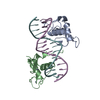
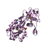
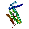
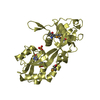
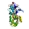
- PDB-1dsz: STRUCTURE OF THE RXR/RAR DNA-BINDING DOMAIN HETERODIMER IN COMPLE... -
+ Open data

- Basic information
| Entry | Database: PDB / ID: 1dsz | ||||||
|---|---|---|---|---|---|---|---|
| Title | STRUCTURE OF THE RXR/RAR DNA-BINDING DOMAIN HETERODIMER IN COMPLEX WITH THE RETINOIC ACID RESPONSE ELEMENT DR1 | ||||||
 Components Components |
| ||||||
 Keywords Keywords | TRANSCRIPTION/DNA / RXR / RAR / nuclear receptor / protein-DNA / retinoic acid / TRANSCRIPTION-DNA COMPLEX | ||||||
| Function / homology |  Function and homology information Function and homology informationSertoli cell fate commitment / positive regulation of binding / trachea cartilage development / glandular epithelial cell development / ventricular cardiac muscle cell differentiation / chondroblast differentiation / embryonic camera-type eye development / growth plate cartilage development / protein kinase B binding / positive regulation of T-helper 2 cell differentiation ...Sertoli cell fate commitment / positive regulation of binding / trachea cartilage development / glandular epithelial cell development / ventricular cardiac muscle cell differentiation / chondroblast differentiation / embryonic camera-type eye development / growth plate cartilage development / protein kinase B binding / positive regulation of T-helper 2 cell differentiation / prostate gland development / negative regulation of granulocyte differentiation / retinoic acid-responsive element binding / negative regulation of cartilage development / NR1H2 & NR1H3 regulate gene expression linked to triglyceride lipolysis in adipose / NR1H2 & NR1H3 regulate gene expression linked to gluconeogenesis / regulation of hematopoietic progenitor cell differentiation / positive regulation of thyroid hormone receptor signaling pathway / NR1H2 & NR1H3 regulate gene expression to limit cholesterol uptake / positive regulation of interleukin-5 production / positive regulation of interleukin-13 production / NR1H2 & NR1H3 regulate gene expression linked to lipogenesis / Carnitine shuttle / retinoic acid binding / outflow tract septum morphogenesis / positive regulation of vitamin D receptor signaling pathway / TGFBR3 expression / response to vitamin A / nuclear vitamin D receptor binding / apoptotic cell clearance / Signaling by Retinoic Acid / heterocyclic compound binding / ureteric bud development / regulation of myelination / DNA-binding transcription repressor activity / NR1H2 & NR1H3 regulate gene expression to control bile acid homeostasis / DNA binding domain binding / positive regulation of lipid metabolic process / limb development / LBD domain binding / positive regulation of interleukin-4 production / positive regulation of lipoprotein transport / face development / nuclear steroid receptor activity / protein kinase A binding / negative regulation of type II interferon production / germ cell development / Synthesis of bile acids and bile salts / alpha-actinin binding / cellular response to estrogen stimulus / negative regulation of tumor necrosis factor production / Synthesis of bile acids and bile salts via 27-hydroxycholesterol / positive regulation of cholesterol efflux / Endogenous sterols / Synthesis of bile acids and bile salts via 7alpha-hydroxycholesterol / monocyte differentiation / response to retinoic acid / cellular response to low-density lipoprotein particle stimulus / positive regulation of bone mineralization / Transcriptional regulation of brown and beige adipocyte differentiation by EBF2 / Recycling of bile acids and salts / retinoic acid receptor signaling pathway / NR1H3 & NR1H2 regulate gene expression linked to cholesterol transport and efflux / cellular response to retinoic acid / Regulation of lipid metabolism by PPARalpha / peptide binding / peroxisome proliferator activated receptor signaling pathway / cell maturation / positive regulation of cell cycle / hormone-mediated signaling pathway / BMAL1:CLOCK,NPAS2 activates circadian expression / response to cytokine / positive regulation of neuron differentiation / RORA,B,C and NR1D1 (REV-ERBA) regulate gene expression / Activation of gene expression by SREBF (SREBP) / Expression of BMAL (ARNTL), CLOCK, and NPAS2 / negative regulation of miRNA transcription / mRNA regulatory element binding translation repressor activity / transcription coregulator binding / RNA polymerase II transcription regulatory region sequence-specific DNA binding / SUMOylation of intracellular receptors / hippocampus development / neural tube closure / Heme signaling / liver development / female pregnancy / PPARA activates gene expression / Transcriptional activation of mitochondrial biogenesis / Cytoprotection by HMOX1 / chromatin DNA binding / Nuclear Receptor transcription pathway / Transcriptional regulation of white adipocyte differentiation / mRNA transcription by RNA polymerase II / regulation of synaptic plasticity / nuclear receptor activity / Activation of anterior HOX genes in hindbrain development during early embryogenesis / multicellular organism growth / transcription coactivator binding / mRNA 5'-UTR binding / RNA polymerase II transcription regulator complex Similarity search - Function | ||||||
| Biological species |  Homo sapiens (human) Homo sapiens (human) | ||||||
| Method |  X-RAY DIFFRACTION / SYNCHROTRON / Resolution: 1.7 Å X-RAY DIFFRACTION / SYNCHROTRON / Resolution: 1.7 Å | ||||||
 Authors Authors | Rastinejad, F. / Wagner, T. / Zhao, Q. / Khorasanizadeh, S. | ||||||
 Citation Citation | Journal: EMBO J. / Year: 2000 Title: Structure of the RXR-RAR DNA-binding complex on the retinoic acid response element DR1. Authors: Rastinejad, F. / Wagner, T. / Zhao, Q. / Khorasanizadeh, S. | ||||||
| History |
|
- Structure visualization
| Structure viewer | Molecule: MolmilJmol/JSmol |
|---|
- Downloads & links
-Download
| PDBx/mmCIF format | 1dsz.cif.gz | 72.5 KB | Display | PDBx/mmCIF format |
|---|---|---|---|---|
| PDB format | pdb1dsz.ent.gz | 49.4 KB | Display | PDB format |
| PDBx/mmJSON format | 1dsz.json.gz | Tree view | PDBx/mmJSON format | |
| Others |  Other downloads Other downloads |
-Validation report
| Arichive directory | https://data.pdbj.org/pub/pdb/validation_reports/ds/1dszftp://data.pdbj.org/pub/pdb/validation_reports/ds/1dsz | HTTPS FTP |
|---|
-Related structure data
| Similar structure data |
|---|


-Links
 PDBj
PDBj


- Assembly
| Deposited unit | 
| ||||||||||
|---|---|---|---|---|---|---|---|---|---|---|---|
| 1 |
| ||||||||||
| Unit cell |
|
-Components
-DNA chain , 2 types, 2 molecules CD
| #1: DNA chain | Mass: 4643.037 Da / Num. of mol.: 1 / Source method: obtained synthetically |
|---|---|
| #2: DNA chain | Mass: 4535.946 Da / Num. of mol.: 1 / Source method: obtained synthetically |
-RETINOIC ACID RECEPTOR ... , 2 types, 2 molecules AB
| #3: Protein | Mass: 10220.006 Da / Num. of mol.: 1 / Fragment: RESIDUES 82-167 Source method: isolated from a genetically manipulated source Source: (gene. exp.) Homo sapiens (human) / Plasmid: PGEX-2T / Production host:  |
|---|---|
| #4: Protein | Mass: 9993.584 Da / Num. of mol.: 1 / Fragment: RESIDUES 129-212 Source method: isolated from a genetically manipulated source Source: (gene. exp.) Homo sapiens (human) / Plasmid: PGEX-2T / Production host: |
-Non-polymers , 2 types, 342 molecules 

| #5: Chemical | ChemComp-ZN /  Mass: 65.409 Da / Num. of mol.: 4 / Source method: obtained synthetically / Formula: Zn Mass: 65.409 Da / Num. of mol.: 4 / Source method: obtained synthetically / Formula: Zn#6: Water | ChemComp-HOH / | Mass: 18.015 Da / Num. of mol.: 338 / Source method: isolated from a natural source / Formula: H2O |
|---|
-Experimental details
-Experiment
| Experiment | Method: X-RAY DIFFRACTION / Number of used crystals: 1 |
|---|
- Sample preparation
| Crystal | Density Matthews: 2.34 Å3/Da / Density % sol: 47.43 % | ||||||||||||||||||||||||||||||||||||||||||||||||
|---|---|---|---|---|---|---|---|---|---|---|---|---|---|---|---|---|---|---|---|---|---|---|---|---|---|---|---|---|---|---|---|---|---|---|---|---|---|---|---|---|---|---|---|---|---|---|---|---|---|
| Crystal grow | Temperature: 281 K / Method: vapor diffusion, hanging drop / pH: 7.5 Details: 18-23% PEG 3350, 25 mM Tris buffer, 5 mM MgCl2, 0.4 M NH4CL, RXR and RAR proteins each at 0.45 mM, DNA duplex at 0.45 mM, pH 7.5, VAPOR DIFFUSION, HANGING DROP, temperature 281K | ||||||||||||||||||||||||||||||||||||||||||||||||
| Components of the solutions |
| ||||||||||||||||||||||||||||||||||||||||||||||||
| Crystal grow | *PLUS Temperature: 8 ℃Details: drop consists of equal volume of protein and reservoir solutions | ||||||||||||||||||||||||||||||||||||||||||||||||
| Components of the solutions | *PLUS
|
-Data collection
| Diffraction | Mean temperature: 113 K |
|---|---|
| Diffraction source | Source: SYNCHROTRON / Site: NSLS  / Beamline: X25 / Wavelength: 0.98 / Beamline: X25 / Wavelength: 0.98 |
| Detector | Type: MARRESEARCH / Detector: AREA DETECTOR / Date: May 11, 1995 |
| Radiation | Protocol: SINGLE WAVELENGTH / Monochromatic (M) / Laue (L): M / Scattering type: x-ray |
| Radiation wavelength | Wavelength: 0.98 Å / Relative weight: 1 |
| Reflection | Resolution: 1.7→50 Å / Num. all: 29834 / Num. obs: 29834 / % possible obs: 94.8 % / Observed criterion σ(I): 0 / Redundancy: 4.1 % / Biso Wilson estimate: 11 Å2 / Rmerge(I) obs: 0.071 / Net I/σ(I): 19.9 |
| Reflection shell | Resolution: 1.7→1.76 Å / Redundancy: 4 % / Rmerge(I) obs: 0.17 / % possible all: 98 |
| Reflection shell | *PLUS % possible obs: 98 % / Mean I/σ(I) obs: 8.8 |
- Processing
| Software |
| |||||||||||||||||||||||||
|---|---|---|---|---|---|---|---|---|---|---|---|---|---|---|---|---|---|---|---|---|---|---|---|---|---|---|
| Refinement | Resolution: 1.7→20 Å / Cross valid method: THROUGHOUT / σ(F): 2 / σ(I): 1 / Stereochemistry target values: X-PLOR
| |||||||||||||||||||||||||
| Refinement step | Cycle: LAST / Resolution: 1.7→20 Å
| |||||||||||||||||||||||||
| Refine LS restraints |
|
