 Movie
Movie Controller

[English] 日本語
 Yorodumi
- PDB-1by4: STRUCTURE AND MECHANISM OF THE HOMODIMERIC ASSEMBLY OF THE RXR ON DNA -
+ Open data

- Basic information
| Entry | Database: PDB / ID: 1by4 | ||||||
|---|---|---|---|---|---|---|---|
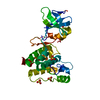
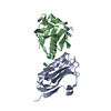
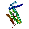
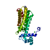
| Title | STRUCTURE AND MECHANISM OF THE HOMODIMERIC ASSEMBLY OF THE RXR ON DNA | ||||||
 Components Components |
| ||||||
 Keywords Keywords | GENE REGULATION/DNA / RXR / NUCLEAR RECEPTOR / HORMONE RESPONSE ELEMENT / PROTEIN-DNA INTERATIONS / GENE REGULATION-DNA COMPLEX | ||||||
| Function / homology |  Function and homology information Function and homology informationretinoic acid-responsive element binding / NR1H2 & NR1H3 regulate gene expression linked to triglyceride lipolysis in adipose / NR1H2 & NR1H3 regulate gene expression linked to gluconeogenesis / positive regulation of thyroid hormone receptor signaling pathway / NR1H2 & NR1H3 regulate gene expression to limit cholesterol uptake / NR1H2 & NR1H3 regulate gene expression linked to lipogenesis / Carnitine shuttle / retinoic acid binding / positive regulation of vitamin D receptor signaling pathway / TGFBR3 expression ...retinoic acid-responsive element binding / NR1H2 & NR1H3 regulate gene expression linked to triglyceride lipolysis in adipose / NR1H2 & NR1H3 regulate gene expression linked to gluconeogenesis / positive regulation of thyroid hormone receptor signaling pathway / NR1H2 & NR1H3 regulate gene expression to limit cholesterol uptake / NR1H2 & NR1H3 regulate gene expression linked to lipogenesis / Carnitine shuttle / retinoic acid binding / positive regulation of vitamin D receptor signaling pathway / TGFBR3 expression / nuclear vitamin D receptor binding / Signaling by Retinoic Acid / NR1H2 & NR1H3 regulate gene expression to control bile acid homeostasis / DNA binding domain binding / positive regulation of lipid metabolic process / nuclear steroid receptor activity / LBD domain binding / positive regulation of lipoprotein transport / Synthesis of bile acids and bile salts / monocyte differentiation / Synthesis of bile acids and bile salts via 27-hydroxycholesterol / positive regulation of cholesterol efflux / Endogenous sterols / Synthesis of bile acids and bile salts via 7alpha-hydroxycholesterol / cellular response to low-density lipoprotein particle stimulus / response to retinoic acid / positive regulation of bone mineralization / retinoic acid receptor signaling pathway / Transcriptional regulation of brown and beige adipocyte differentiation by EBF2 / Recycling of bile acids and salts / NR1H3 & NR1H2 regulate gene expression linked to cholesterol transport and efflux / peroxisome proliferator activated receptor signaling pathway / hormone-mediated signaling pathway / Regulation of lipid metabolism by PPARalpha / cell maturation / peptide binding / BMAL1:CLOCK,NPAS2 activates circadian expression / RORA,B,C and NR1D1 (REV-ERBA) regulate gene expression / Activation of gene expression by SREBF (SREBP) / Expression of BMAL (ARNTL), CLOCK, and NPAS2 / transcription coregulator binding / SUMOylation of intracellular receptors / RNA polymerase II transcription regulatory region sequence-specific DNA binding / Heme signaling / PPARA activates gene expression / Transcriptional activation of mitochondrial biogenesis / Cytoprotection by HMOX1 / Nuclear Receptor transcription pathway / Transcriptional regulation of white adipocyte differentiation / mRNA transcription by RNA polymerase II / nuclear receptor activity / RNA polymerase II transcription regulator complex / Activation of anterior HOX genes in hindbrain development during early embryogenesis / Transcriptional regulation of granulopoiesis / sequence-specific double-stranded DNA binding / nervous system development / MLL4 and MLL3 complexes regulate expression of PPARG target genes in adipogenesis and hepatic steatosis / double-stranded DNA binding / transcription regulator complex / sequence-specific DNA binding / DNA-binding transcription factor activity, RNA polymerase II-specific / cell differentiation / signaling receptor complex / transcription cis-regulatory region binding / RNA polymerase II cis-regulatory region sequence-specific DNA binding / DNA-binding transcription factor activity / positive regulation of DNA-templated transcription / chromatin / enzyme binding / negative regulation of transcription by RNA polymerase II / positive regulation of transcription by RNA polymerase II / mitochondrion / zinc ion binding / nucleoplasm / identical protein binding / nucleus / cytosol Similarity search - Function | ||||||
| Biological species |  Homo sapiens (human) Homo sapiens (human) | ||||||
| Method |  X-RAY DIFFRACTION / SYNCHROTRON / MOLECULAR REPLACEMENT / Resolution: 2.1 Å X-RAY DIFFRACTION / SYNCHROTRON / MOLECULAR REPLACEMENT / Resolution: 2.1 Å | ||||||
 Authors Authors | Zhao, Q. / Chasse, S.A. / Devarakonda, S. / Sierk, M.L. / Ahvazi, B. / Sigler, P.B. / Rastinejad, F. | ||||||
 Citation Citation | Journal: J.Mol.Biol. / Year: 2000 Title: Structural basis of RXR-DNA interactions. Authors: Zhao, Q. / Chasse, S.A. / Devarakonda, S. / Sierk, M.L. / Ahvazi, B. / Rastinejad, F. | ||||||
| History |
|
- Structure visualization
| Structure viewer | Molecule: MolmilJmol/JSmol |
|---|
- Downloads & links
-Download
| PDBx/mmCIF format | 1by4.cif.gz | 121.1 KB | Display | PDBx/mmCIF format |
|---|---|---|---|---|
| PDB format | pdb1by4.ent.gz | 88.1 KB | Display | PDB format |
| PDBx/mmJSON format | 1by4.json.gz | Tree view | PDBx/mmJSON format | |
| Others |  Other downloads Other downloads |
-Validation report
| Arichive directory | https://data.pdbj.org/pub/pdb/validation_reports/by/1by4ftp://data.pdbj.org/pub/pdb/validation_reports/by/1by4 | HTTPS FTP |
|---|
-Related structure data
| Related structure data |  2nulS S: Starting model for refinement |
|---|---|
| Similar structure data |


-Links
 PDBj
PDBj


- Assembly
| Deposited unit | 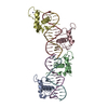
| ||||||||||
|---|---|---|---|---|---|---|---|---|---|---|---|
| 1 | 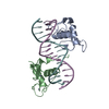
| ||||||||||
| 2 | 
| ||||||||||
| Unit cell |
|
-Components
| #1: DNA chain | Mass: 4947.229 Da / Num. of mol.: 2 / Source method: obtained synthetically #2: DNA chain | Mass: 4519.947 Da / Num. of mol.: 2 / Source method: obtained synthetically #3: Protein | Mass: 9651.209 Da / Num. of mol.: 4 Source method: isolated from a genetically manipulated source Source: (gene. exp.) Homo sapiens (human) / Production host:  #4: Chemical | ChemComp-ZN /   Mass: 65.409 Da / Num. of mol.: 8 / Source method: obtained synthetically / Formula: Zn Mass: 65.409 Da / Num. of mol.: 8 / Source method: obtained synthetically / Formula: Zn#5: Water | ChemComp-HOH / |  Mass: 18.015 Da / Num. of mol.: 230 / Source method: isolated from a natural source / Formula: H2O Mass: 18.015 Da / Num. of mol.: 230 / Source method: isolated from a natural source / Formula: H2O |
|---|
-Experimental details
-Experiment
| Experiment | Method: X-RAY DIFFRACTION / Number of used crystals: 1 |
|---|
- Sample preparation
| Crystal | Density Matthews: 2.9 Å3/Da / Density % sol: 47 % | ||||||||||||||||||||||||||||||||||||||||||||||||
|---|---|---|---|---|---|---|---|---|---|---|---|---|---|---|---|---|---|---|---|---|---|---|---|---|---|---|---|---|---|---|---|---|---|---|---|---|---|---|---|---|---|---|---|---|---|---|---|---|---|
| Crystal grow | pH: 7.5 Details: 25 MM TRIS BUFFER (PH 7.5),20% PEG3350, 5 MM MGCL2, 400MM NH4CL | ||||||||||||||||||||||||||||||||||||||||||||||||
| Crystal grow | *PLUS Temperature: 8 ℃ / Method: vapor diffusion, hanging drop | ||||||||||||||||||||||||||||||||||||||||||||||||
| Components of the solutions | *PLUS
|
-Data collection
| Diffraction | Mean temperature: 113 K |
|---|---|
| Diffraction source | Source: SYNCHROTRON / Site: EMBL/DESY, HAMBURG  / Beamline: X11 / Wavelength: 0.9095 / Beamline: X11 / Wavelength: 0.9095 |
| Detector | Type: MARRESEARCH / Detector: IMAGE PLATE / Date: Dec 14, 1997 |
| Radiation | Protocol: SINGLE WAVELENGTH / Monochromatic (M) / Laue (L): M / Scattering type: x-ray |
| Radiation wavelength | Wavelength: 0.9095 Å / Relative weight: 1 |
| Reflection | Resolution: 2.1→6 Å / Num. obs: 46828 / % possible obs: 85.7 % / Observed criterion σ(I): 2 / Redundancy: 1.8 % / Rmerge(I) obs: 0.081 / Net I/σ(I): 10.39 |
| Reflection shell | Resolution: 2.1→2.18 Å / Rmerge(I) obs: 0.419 / % possible all: 83.2 |
| Reflection | *PLUS Lowest resolution: 6 Å |
| Reflection shell | *PLUS Highest resolution: 2.1 Å / % possible obs: 83.2 % |
- Processing
| Software |
| ||||||||||||||||||||||||||||||||||||||||||||||||||||||||||||
|---|---|---|---|---|---|---|---|---|---|---|---|---|---|---|---|---|---|---|---|---|---|---|---|---|---|---|---|---|---|---|---|---|---|---|---|---|---|---|---|---|---|---|---|---|---|---|---|---|---|---|---|---|---|---|---|---|---|---|---|---|---|
| Refinement | Method to determine structure: MOLECULAR REPLACEMENT Starting model: 2NUL Resolution: 2.1→10 Å / Data cutoff high absF: 10000000 / Data cutoff low absF: 0.001 / σ(F): 2 / Details: extra atom O2* on DC 2494 of chain G was removed.
| ||||||||||||||||||||||||||||||||||||||||||||||||||||||||||||
| Refinement step | Cycle: LAST / Resolution: 2.1→10 Å
| ||||||||||||||||||||||||||||||||||||||||||||||||||||||||||||
| Refine LS restraints |
| ||||||||||||||||||||||||||||||||||||||||||||||||||||||||||||
| LS refinement shell | Resolution: 2.1→2.19 Å / Total num. of bins used: 8 /
| ||||||||||||||||||||||||||||||||||||||||||||||||||||||||||||
| Xplor file |
| ||||||||||||||||||||||||||||||||||||||||||||||||||||||||||||
| Software | *PLUS Name: X-PLOR / Version: 3.851 / Classification: refinement | ||||||||||||||||||||||||||||||||||||||||||||||||||||||||||||
| Refinement | *PLUS Highest resolution: 2.1 Å / Lowest resolution: 10 Å / σ(F): 2 / % reflection Rfree: 5 % / Rfactor obs: 0.219 / Rfactor Rfree: 0.288 | ||||||||||||||||||||||||||||||||||||||||||||||||||||||||||||
| Solvent computation | *PLUS | ||||||||||||||||||||||||||||||||||||||||||||||||||||||||||||
| Displacement parameters | *PLUS | ||||||||||||||||||||||||||||||||||||||||||||||||||||||||||||
| Refine LS restraints | *PLUS
| ||||||||||||||||||||||||||||||||||||||||||||||||||||||||||||
| LS refinement shell | *PLUS Rfactor Rfree: 0.352 / Rfactor Rwork: 0.345 |
