 Movie
Movie Controller


+ Open data

- Basic information
| Entry | Database: PDB / ID: 3ub2 | ||||||
|---|---|---|---|---|---|---|---|
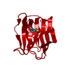
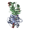
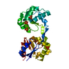
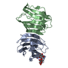
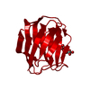
| Title | TIR domain of Mal/TIRAP | ||||||
 Components Components | Toll/interleukin-1 receptor domain-containing adapter protein | ||||||
 Keywords Keywords | IMMUNE SYSTEM / TIR domain / TLRs adaptor | ||||||
| Function / homology |  Function and homology information Function and homology informationpositive regulation of interleukin-15 production / TIRAP-dependent toll-like receptor 4 signaling pathway / toll-like receptor TLR1:TLR2 signaling pathway / cellular response to bacterial lipopeptide / regulation of interferon-beta production / positive regulation of toll-like receptor 3 signaling pathway / Toll-like receptor 4 binding / positive regulation of toll-like receptor 2 signaling pathway / Toll-like receptor 2 binding / positive regulation of chemokine (C-X-C motif) ligand 1 production ...positive regulation of interleukin-15 production / TIRAP-dependent toll-like receptor 4 signaling pathway / toll-like receptor TLR1:TLR2 signaling pathway / cellular response to bacterial lipopeptide / regulation of interferon-beta production / positive regulation of toll-like receptor 3 signaling pathway / Toll-like receptor 4 binding / positive regulation of toll-like receptor 2 signaling pathway / Toll-like receptor 2 binding / positive regulation of chemokine (C-X-C motif) ligand 1 production / positive regulation of toll-like receptor 4 signaling pathway / myeloid cell differentiation / MyD88 deficiency (TLR2/4) / positive regulation of chemokine (C-X-C motif) ligand 2 production / positive regulation of neutrophil chemotaxis / 3'-UTR-mediated mRNA stabilization / IRAK4 deficiency (TLR2/4) / toll-like receptor 4 signaling pathway / MyD88:MAL(TIRAP) cascade initiated on plasma membrane / MyD88-dependent toll-like receptor signaling pathway / extrinsic component of cytoplasmic side of plasma membrane / regulation of innate immune response / cellular response to lipoteichoic acid / endocytic vesicle / positive regulation of interleukin-12 production / positive regulation of B cell proliferation / phosphatidylinositol-4,5-bisphosphate binding / signaling adaptor activity / protein kinase C binding / positive regulation of interleukin-8 production / positive regulation of protein-containing complex assembly / positive regulation of JNK cascade / positive regulation of interleukin-6 production / ruffle membrane / positive regulation of tumor necrosis factor production / ER-Phagosome pathway / response to lipopolysaccharide / molecular adaptor activity / positive regulation of ERK1 and ERK2 cascade / protein-macromolecule adaptor activity / positive regulation of canonical NF-kappaB signal transduction / cell surface receptor signaling pathway / defense response to Gram-positive bacterium / inflammatory response / innate immune response / cell surface / identical protein binding / plasma membrane / cytosol / cytoplasm Similarity search - Function | ||||||
| Biological species |  Homo sapiens (human) Homo sapiens (human) | ||||||
| Method |  X-RAY DIFFRACTION / SYNCHROTRON / SAD / Resolution: 2.4 Å X-RAY DIFFRACTION / SYNCHROTRON / SAD / Resolution: 2.4 Å | ||||||
 Authors Authors | Shen, Y. / Lin, Z. | ||||||
 Citation Citation | Journal: Plos One / Year: 2012 Title: Structural Insights into TIR Domain Specificity of the Bridging Adaptor Mal in TLR4 Signaling Authors: Lin, Z. / Lu, J. / Zhou, W. / Shen, Y. | ||||||
| History |
|
- Structure visualization
| Structure viewer | Molecule: MolmilJmol/JSmol |
|---|
- Downloads & links
-Download
| PDBx/mmCIF format | 3ub2.cif.gz | 37.9 KB | Display | PDBx/mmCIF format |
|---|---|---|---|---|
| PDB format | pdb3ub2.ent.gz | 25.3 KB | Display | PDB format |
| PDBx/mmJSON format | 3ub2.json.gz | Tree view | PDBx/mmJSON format | |
| Others |  Other downloads Other downloads |
-Validation report
| Arichive directory | https://data.pdbj.org/pub/pdb/validation_reports/ub/3ub2ftp://data.pdbj.org/pub/pdb/validation_reports/ub/3ub2 | HTTPS FTP |
|---|
-Related structure data


-Links
 PDBj
PDBj


- Assembly
| Deposited unit | 
| ||||||||
|---|---|---|---|---|---|---|---|---|---|
| 1 | 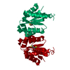
| ||||||||
| Unit cell |
|
-Components
| #1: Protein | Mass: 15970.159 Da / Num. of mol.: 1 / Fragment: TIR domain, UNP residues 78-221 Source method: isolated from a genetically manipulated source Source: (gene. exp.) Homo sapiens (human) / Gene: TIRAP, MAL / Production host:  |
|---|---|
| #2: Chemical | ChemComp-DTT /   Mass: 154.251 Da / Num. of mol.: 1 / Source method: obtained synthetically / Formula: C4H10O2S2 Mass: 154.251 Da / Num. of mol.: 1 / Source method: obtained synthetically / Formula: C4H10O2S2 |
| #3: Water | ChemComp-HOH /  Mass: 18.015 Da / Num. of mol.: 11 / Source method: isolated from a natural source / Formula: H2O Mass: 18.015 Da / Num. of mol.: 11 / Source method: isolated from a natural source / Formula: H2O |
| Has protein modification | Y |
-Experimental details
-Experiment
| Experiment | Method: X-RAY DIFFRACTION / Number of used crystals: 2 |
|---|
- Sample preparation
| Crystal | Density Matthews: 4.9 Å3/Da / Density % sol: 74.88 % |
|---|---|
| Crystal grow | Temperature: 298 K / Method: vapor diffusion, hanging drop / pH: 9.5 Details: 0.1M CHES, 12-20% Glycerol, 150-300mM sodium chloride, 10mM DTT, pH 9.5, VAPOR DIFFUSION, HANGING DROP, temperature 298.0K |
-Data collection
| Diffraction | Mean temperature: 100 K |
|---|---|
| Diffraction source | Source: SYNCHROTRON / Site: SSRF  / Beamline: BL17U / Wavelength: 0.9795 Å / Beamline: BL17U / Wavelength: 0.9795 Å |
| Detector | Type: ADSC QUANTUM 315r / Detector: CCD / Date: Jun 25, 2011 |
| Radiation | Monochromator: Si 111 CHANNEL / Protocol: SINGLE WAVELENGTH / Monochromatic (M) / Laue (L): M / Scattering type: x-ray |
| Radiation wavelength | Wavelength: 0.9795 Å / Relative weight: 1 |
| Reflection | Resolution: 2.4→50 Å / Num. obs: 12504 / % possible obs: 87.8 % / Observed criterion σ(F): 2 / Observed criterion σ(I): 2 / Biso Wilson estimate: 55 Å2 |
| Reflection shell | Resolution: 2.4→2.49 Å / % possible all: 80 |
- Processing
| Software |
| ||||||||||||||||||||||||||||||||||||
|---|---|---|---|---|---|---|---|---|---|---|---|---|---|---|---|---|---|---|---|---|---|---|---|---|---|---|---|---|---|---|---|---|---|---|---|---|---|
| Refinement | Method to determine structure: SAD / Resolution: 2.4→39.14 Å / Rfactor Rfree error: 0.01 / Data cutoff high absF: 1011124.76 / Data cutoff low absF: 0 / Isotropic thermal model: RESTRAINED / Cross valid method: THROUGHOUT / σ(F): 2 / Stereochemistry target values: Engh & Huber / Details: BULK SOLVENT MODEL USED
| ||||||||||||||||||||||||||||||||||||
| Solvent computation | Solvent model: FLAT MODEL / Bsol: 53.23 Å2 / ksol: 0.4 e/Å3 | ||||||||||||||||||||||||||||||||||||
| Displacement parameters | Biso mean: 61.8 Å2
| ||||||||||||||||||||||||||||||||||||
| Refine analyze |
| ||||||||||||||||||||||||||||||||||||
| Refinement step | Cycle: LAST / Resolution: 2.4→39.14 Å
| ||||||||||||||||||||||||||||||||||||
| Refine LS restraints |
| ||||||||||||||||||||||||||||||||||||
| LS refinement shell | Resolution: 2.4→2.55 Å / Rfactor Rfree error: 0.038 / Total num. of bins used: 6
| ||||||||||||||||||||||||||||||||||||
| Xplor file |
|
